We’re excited to share the latest edition of SEM nanoNews with you! You’ll find updates on Phenom software, new additions to our website, and everything you need to know about upcoming events, webinars, blogs, and our expanded image gallery—all centered around Scanning Electron Microscopy (SEM).
Don’t miss out- Follow us on LinkedIn to stay up to date on news, publications and more!
NEWS & UPDATES
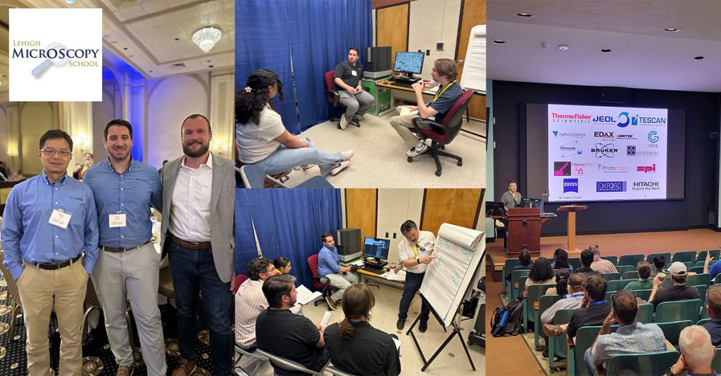
We had a fantastic time at the Lehigh Microscopy School last month!
Dr. Jining Xie, William K. Podrazky, and Dr. Vincent Pastore, led hands-on training sessions with the Phenom XL Benchtop SEM, and it was clear that many attendees were genuinely surprised by what this compact system can do.
We fielded a lot of great questions, and these came up again and again:
Can it actually do EDS?
Yes! The Phenoms can be equipped with a Silicon Drift Detector (SDD), offering fast, accurate elemental analysis. The integrated software makes it easy to collect point spectra, line scans, and elemental maps, and allows automation of EDS analysis tasks.
This is just a teaching tool, right? Can it even collect publication-quality images?
It is not just for teaching! While they are easy to set up and use, the Phenoms are routinely used in some of the most cutting-edge research labs around the world. Phenom micrographs have been published in many journals and have also won image contests.
It’s always rewarding to experience the excitement generated by the Phenom SEMs and see skepticism turned into enthusiasm!
CONFERENCE CORNER
UPCOMING EVENTS:
Microscopy & Microanalysis 2025

Join us in Salt Lake City, Utah for the highlight of the microscopy calendar – M&M 2025! Nanoscience Instruments is back for this annual summertime conference, and this time we’re showcasing in Booth #1925.

ON-DEMAND WEBINARS
Using Automated SEM/EDS Analysis to Enhance Parts Cleanliness
Maintaining technical cleanliness is critical in industries where contamination can compromise product performance, safety, and compliance. From automotive and aerospace to medical devices and microelectronics, stringent cleanliness standards help mitigate …
Superfast Imaging for Electron Microscopy: Up to 100x Faster, 100x Less Data and 100x Less Dose
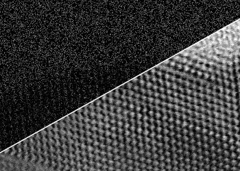
In this webinar, we will introduce ‘Compressed Sensing’ software from SenseAI, which samples a fraction of the data without loss of any inherent information. This generates images faster with significantly …
Applications for Desktop SEM in Geology
Scanning Electron Microscopy (SEM) has become a vital tool in geology, offering high-resolution imaging and elemental analysis for understanding the composition, texture, and geologic history of rocks, minerals, and fossils. …
BLOGS
Automated SEM: The Future of Particle Analysis
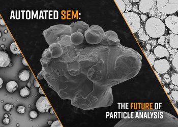
Particle analysis involves characterizing the size, morphology, and composition of powders and particulate matter to describe their properties in a precise and statistically significant manner. Some of the most common …
Benchtop STEM-in-SEM: A Powerful Tool for Tissue Ultrastructure Studies
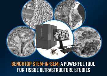
Understanding the ultrastructure of biological tissues is essential for advancing medical research, disease diagnostics, and drug development. Traditionally, histological techniques such as optical microscopy have been the standard for tissue …
Enhancing SEM Performance with Ion Milling Technology
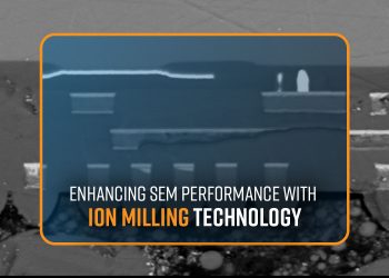
In material science, cross-section polishing is a critical method to enable the detailed examination of a sample material’s microstructure. This technique is essential for understanding the composition, properties, and potential …
Desktop Scanning Electron Microscopy in Geology
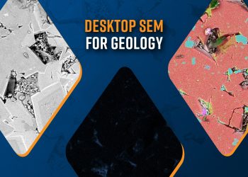
Scanning Electron Microscopy (SEM) is a versatile tool in geology used in high-resolution imaging and chemical analysis of rocks, minerals, and microfossils. SEM provides detailed images of mineral morphology and …