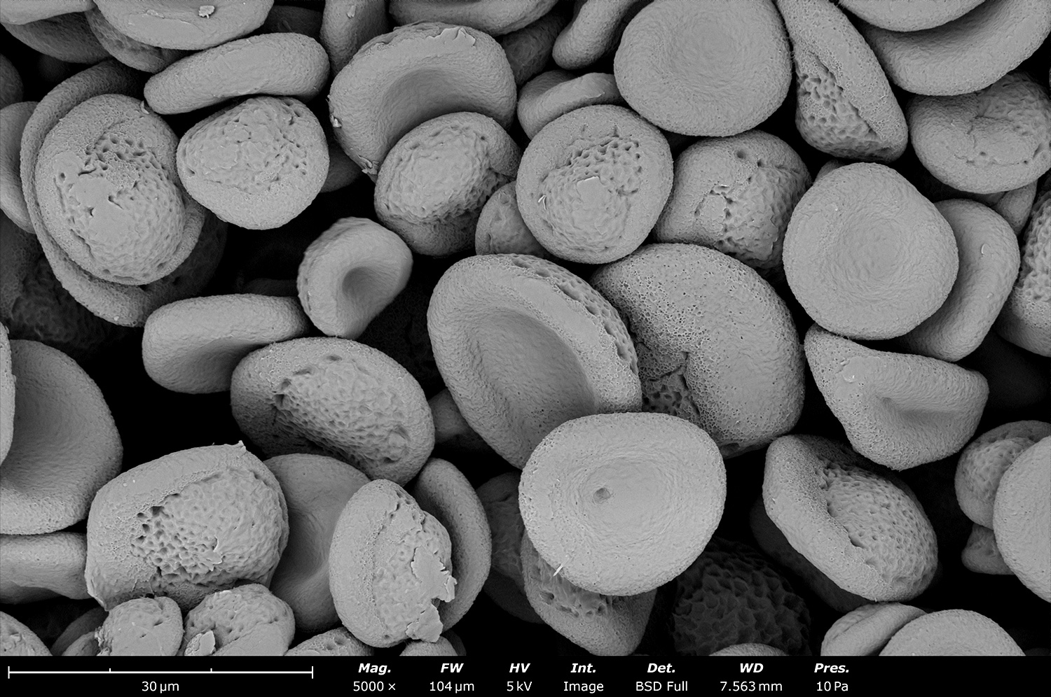
Electron micrograph of electrosprayed polycaprolactone (PCL) microparticles made with a Fluidnatek LE-50. These particles were made with a high boiling point and low vapor pressure solvent allowing them to obtain a smooth and rounded morphology. After collecting onto a liquid reservoir, the media was evaporated, and the PCL particles agglomerated for future experiments
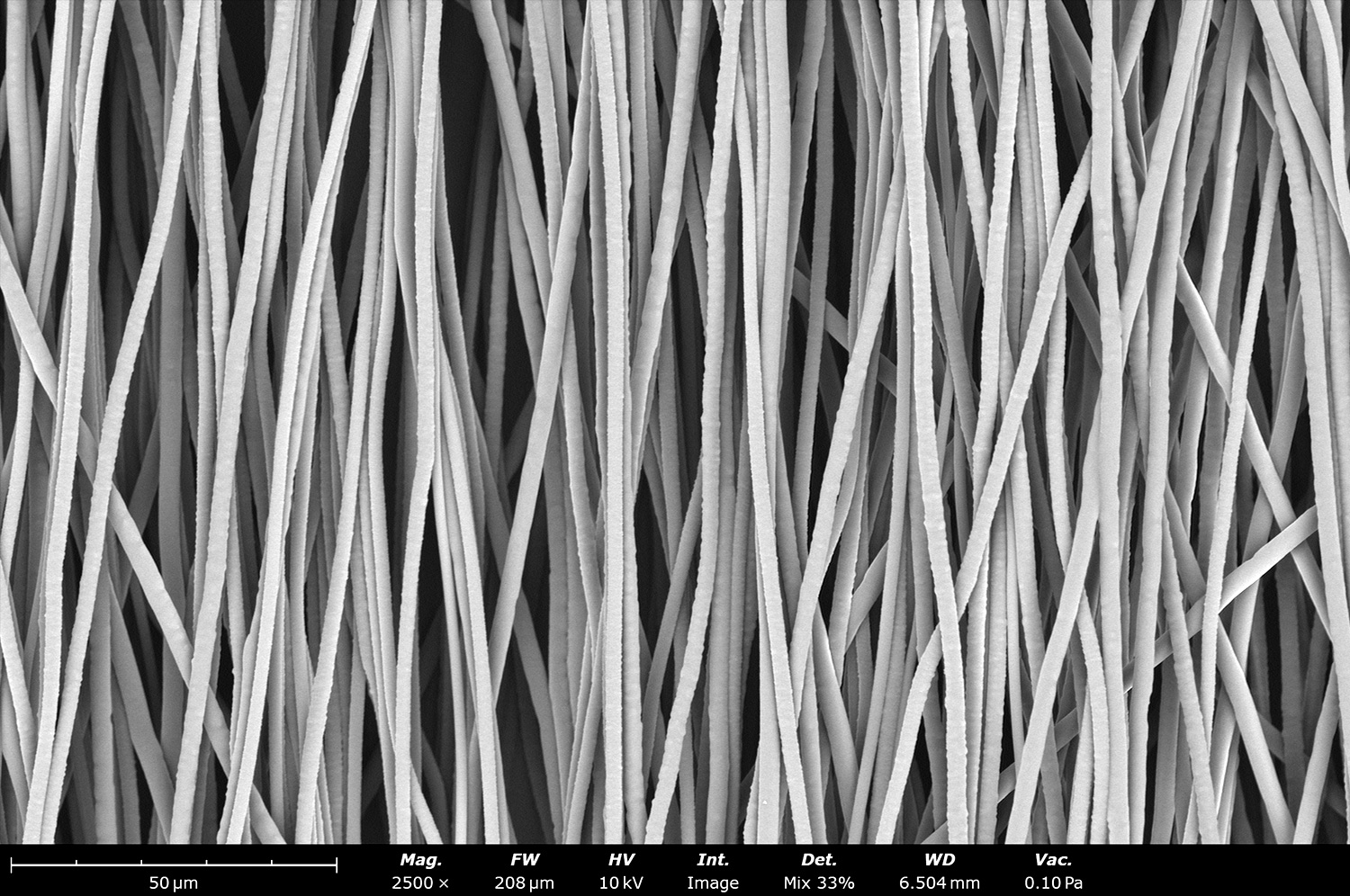
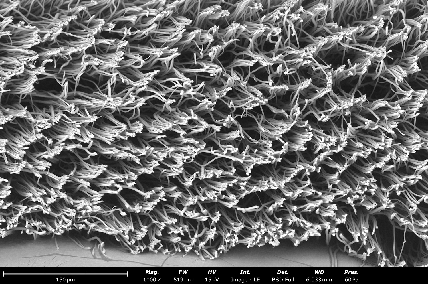
Polycaprolactone (PCL) is a common synthetic polymer used for tissue engineering, medical devices and wound healing applications. This image shows PCL microfibers made with dichloromethane as the solvent. The resultant porous structure, due to rapid solvent evaporation, is desired to increase surface roughness and promote cell proliferation.
Co-spinning offers multiple advantages for sample development including improving mechanical properties, covering large pores with nanoscale fibers, having each fiber with a unique active pharmaceutical ingredient (API), or a drug, and maximize area density. Co-processed samples can be made of two different fibers, two different particles, or a combination of fibers and particles.
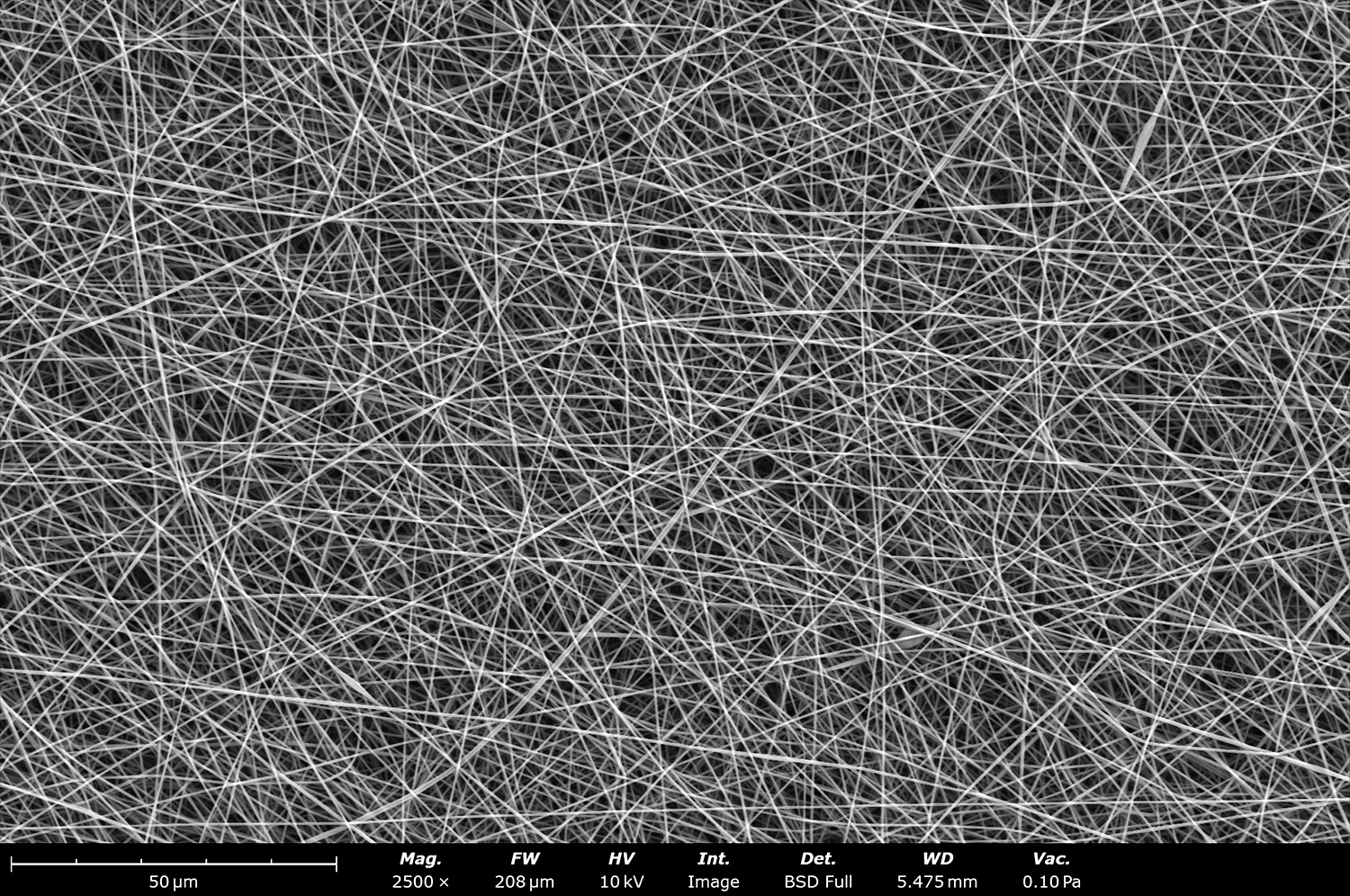
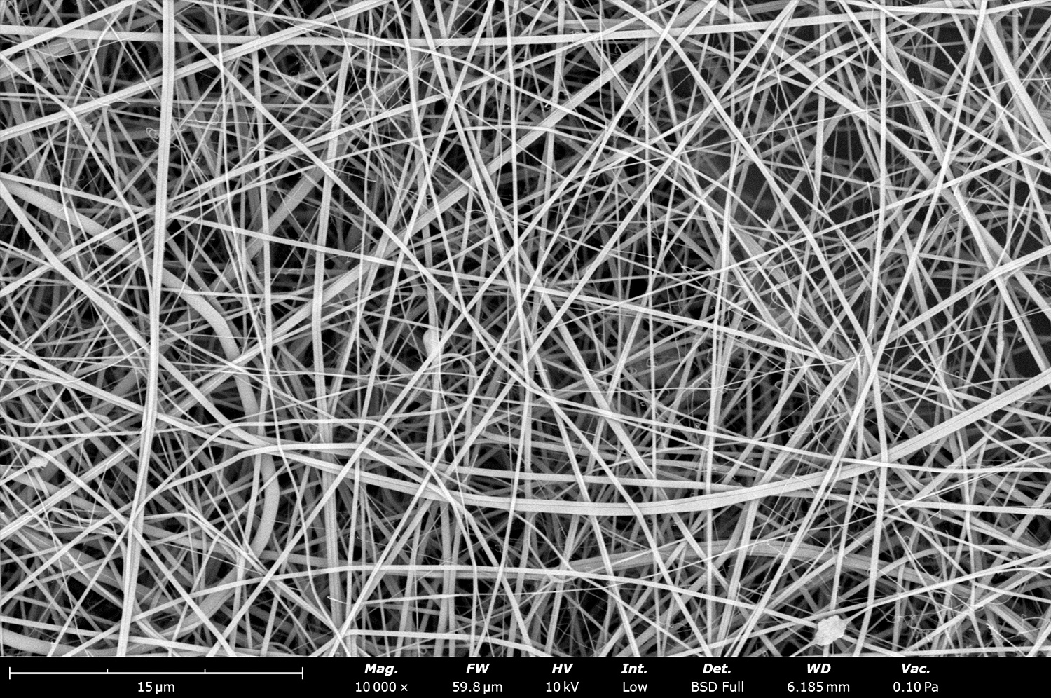
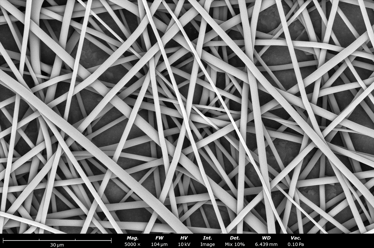
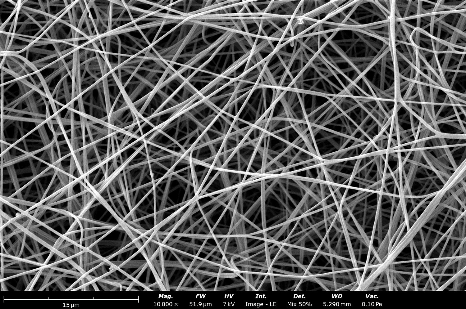
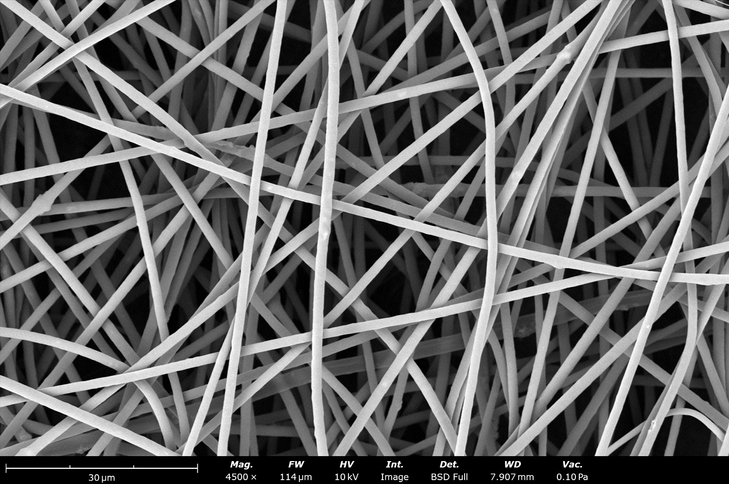
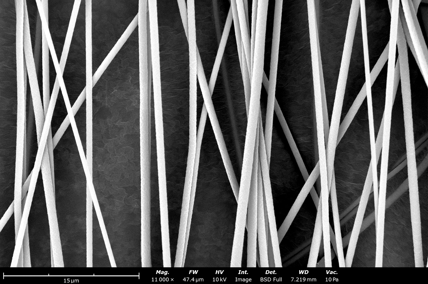
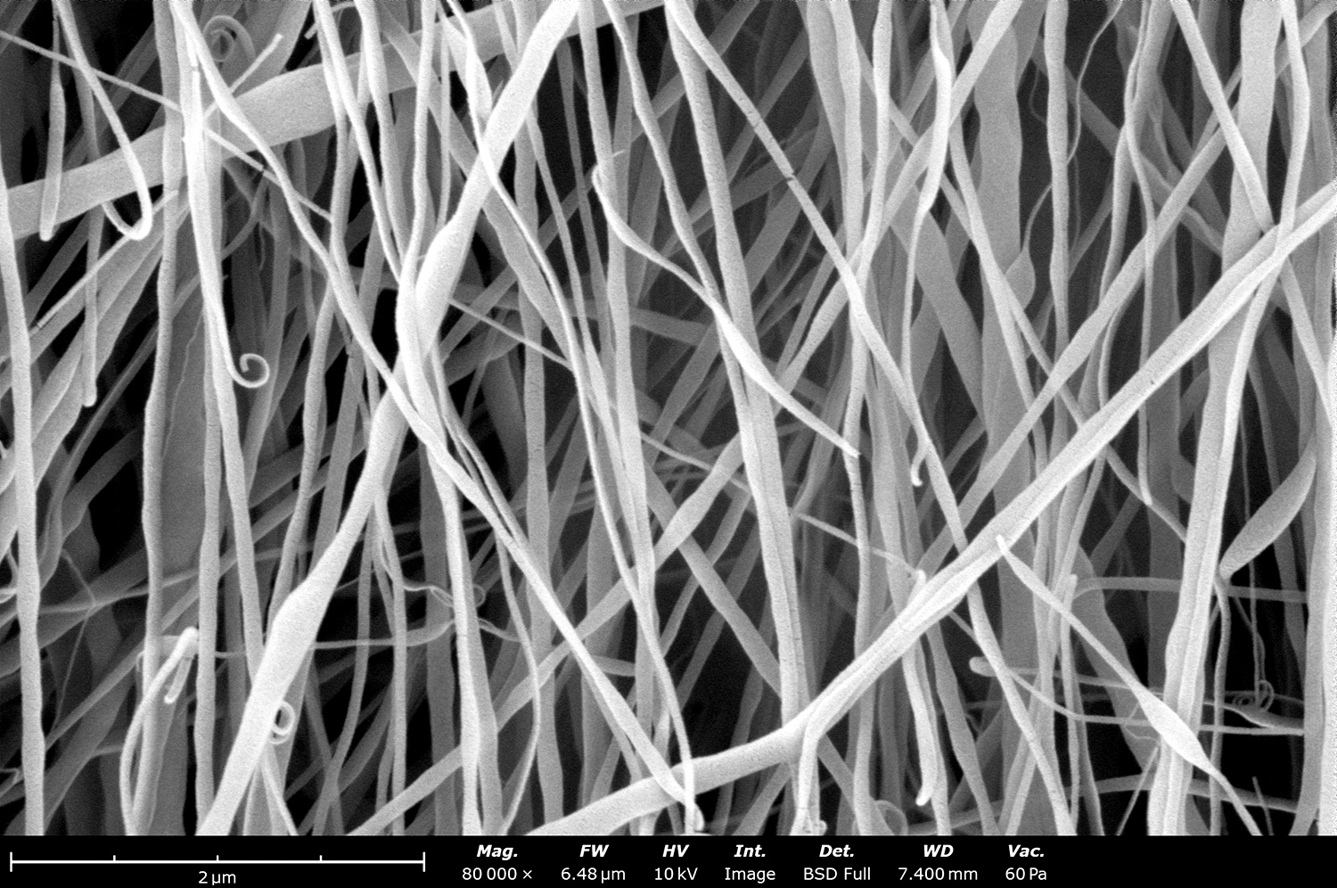
Closer look at electrospun polyacrylonitrile (PAN) fibers made using the Fluidnatek LE-50. These PAN fibers have a standard deviation less than 50 nm as they were developed under tight environmental conditions. These results can be obtained all year round when temperature and relative humidity and tightly controlled with the Fluidnatek technology.
Electron micrograph of electrospun polyacrylonitrile (PAN) made using the Fluidnatek LE-100. These fibers were collected on top of a polyethylene with carbon black substrate to allow for easy sample removal. PAN fibers are typically used for filtration, energy storage applications and to coat medical devices.
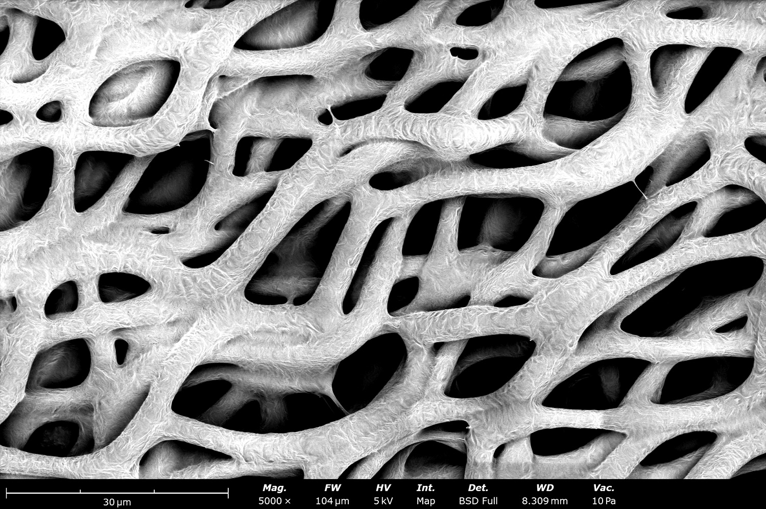
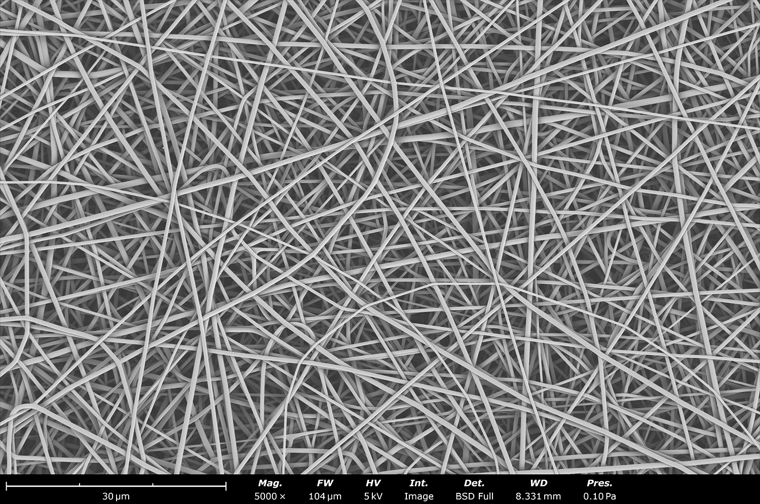
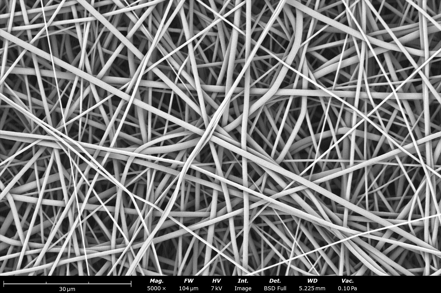
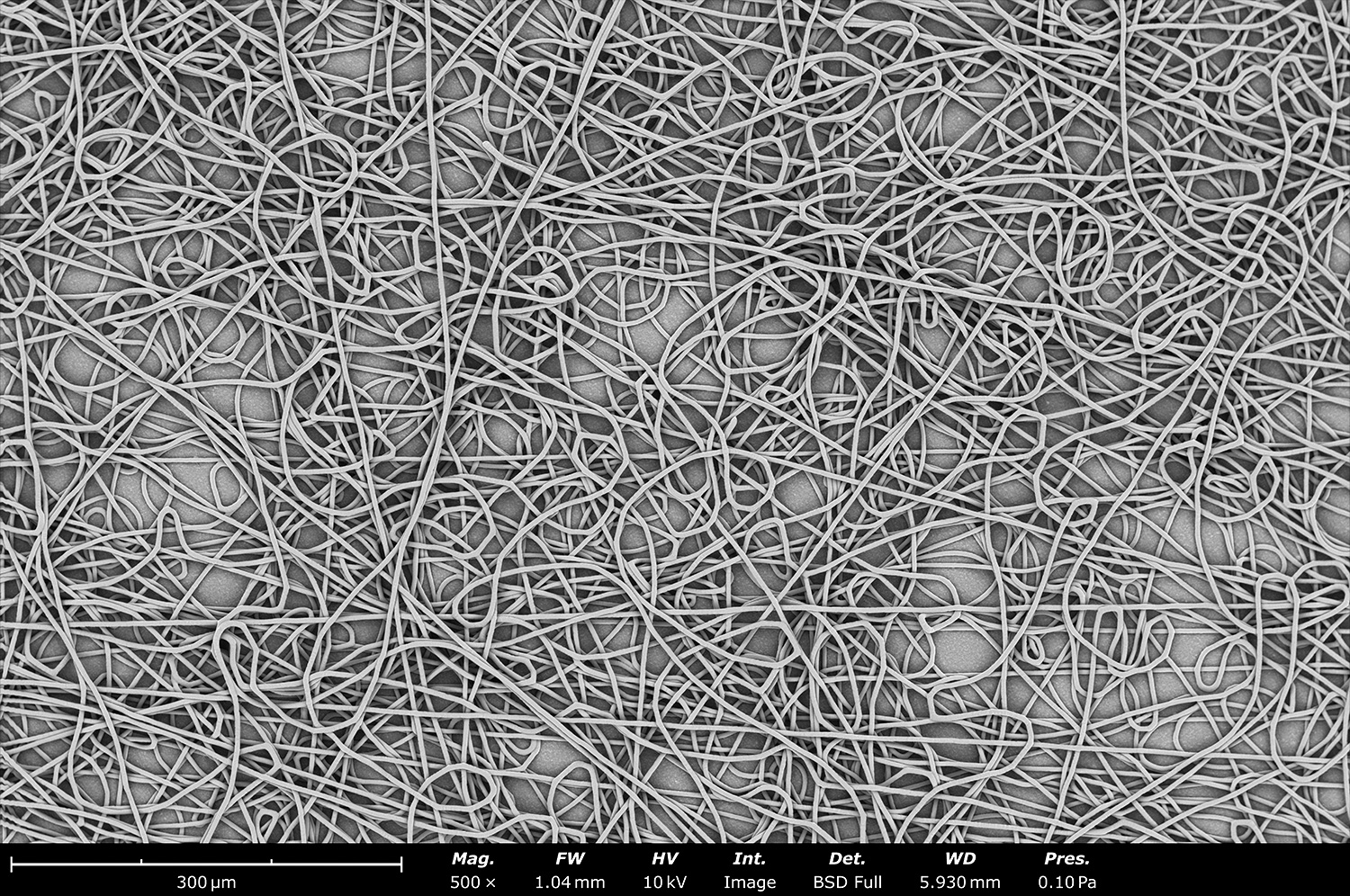
Backscattered electron micrograph of electrospun polycaprolactone (PCL) made using the Fluidnatek LE-500. To achieve reproducibility and fiber consistency, the electrospinning conditions were set to 24 ± 1°C and 40 ± 3%. These randomly oriented fibers offer a pore size around 20 µm to allow for cell infiltration for tissue engineering related applications.
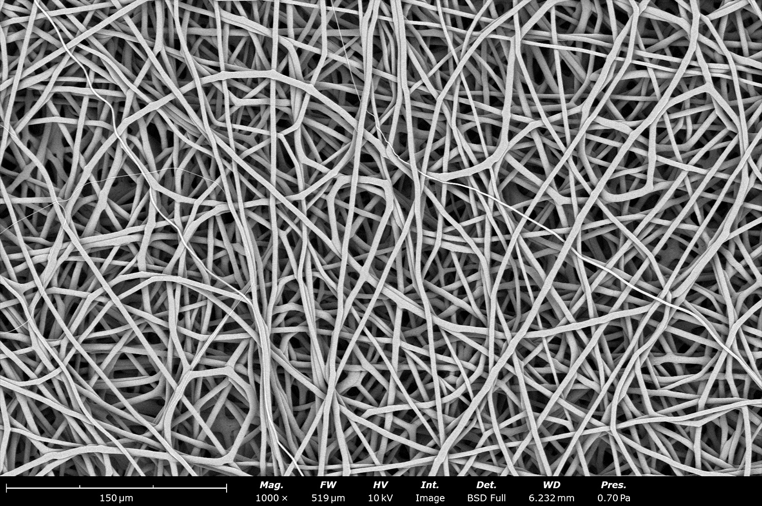
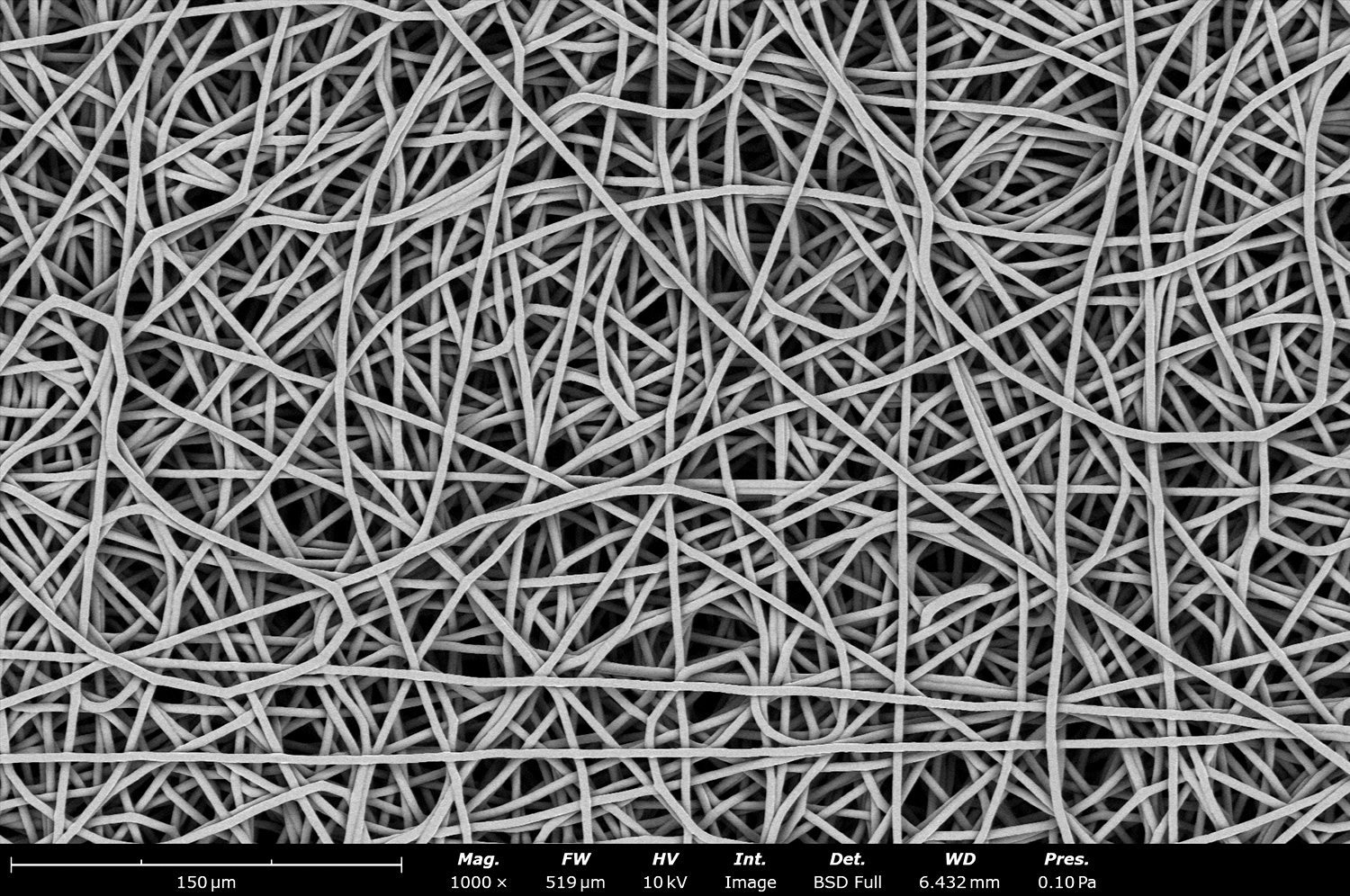
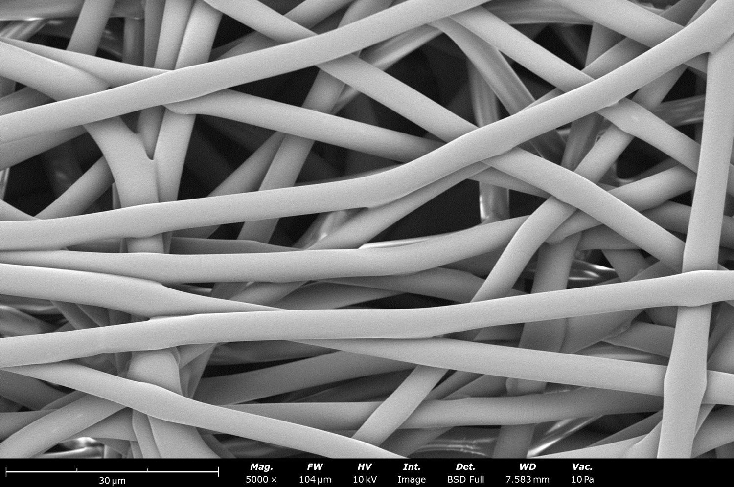
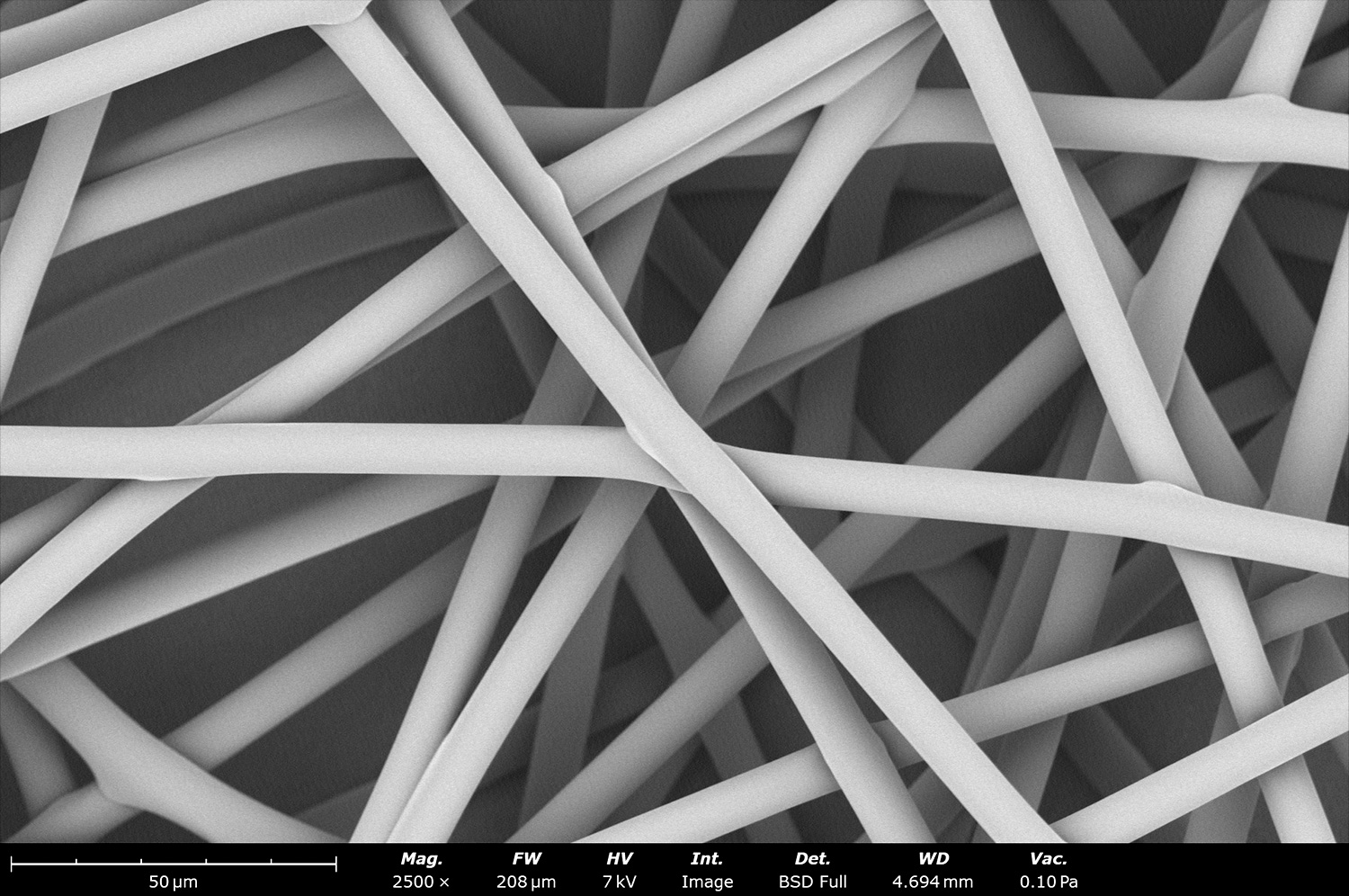
This SEM image shows electrospun polycaprolactone (PCL) made in a Fluidnatek LE-50 at 1,000 rpm and at a translational needle speed of 20 mm/s and a low voltage of -4 kV. The combination of slow translational speed, along with the high rpm and low voltage, allowed the fibers to be collected with a peculiar wavy pattern with significant amount of fiber-fiber bonding that can increase mechanical properties of the final sample.
Microstructure of an electrospun nerve conduit made out of electrospun polylactic acid (PLA) made in the Fluidnatek LE-100. Characteristic features of this sample include the perpendicular aligned PLA fibers on the central portion of the sample (right side of the image) and randomly oriented PLA fibers surrounding the aligned fibers. The packing density of the aligned layers can be controlled with the Fluidnatek technology to fine tune final sample.
Micrograph of electrospun polybutylene succinate (PBS), a biodegradable polymer with water and carbon dioxide byproducts upon degradation by microorganisms. These plant based fibers were electrospun with the Fluidnatek LE-50 and are currently being used for single use food packaging and related applications.
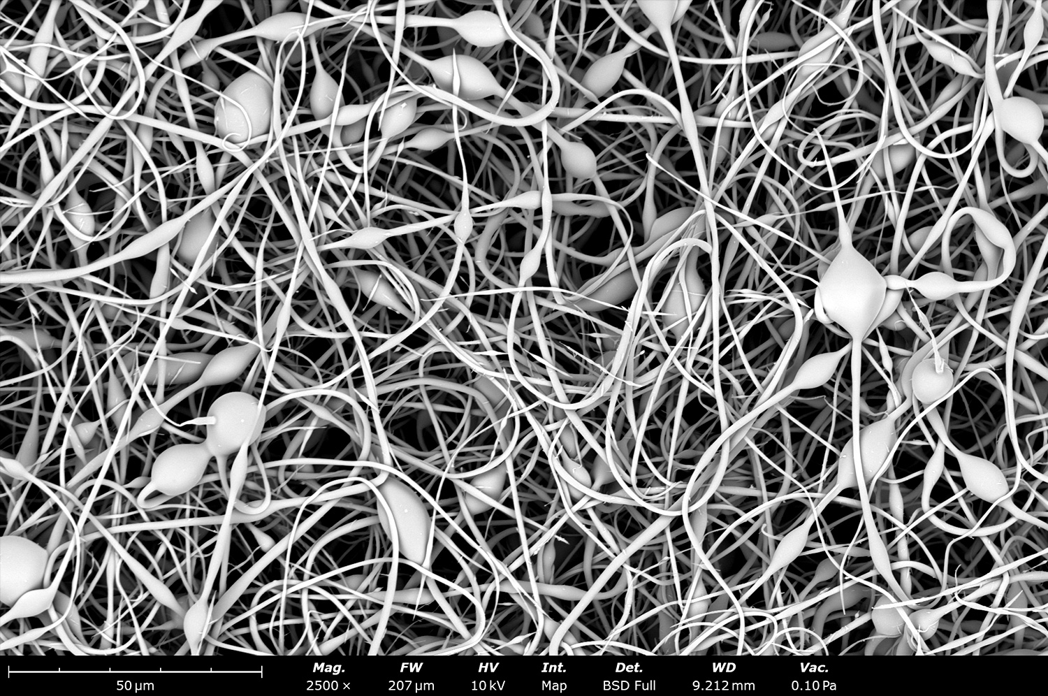
Electron micrograph of biodegradable beaded fibers made out of polycaprolactone (PCL) using the Fluidnatek LE-100 with a 5 mm outside diameter mandrel rotating at 200 rpm. This image was acquired by mixing the signal of the backscatter and secondary electron detectors in a 1:1 ratio. In the past, this type of fiber morphology was thought to be a disadvantage in the electrospinning field. These days the beaded fiber structure is used for drug encapsulation, coat medical devices, increase efficiency of air filtration, among others.
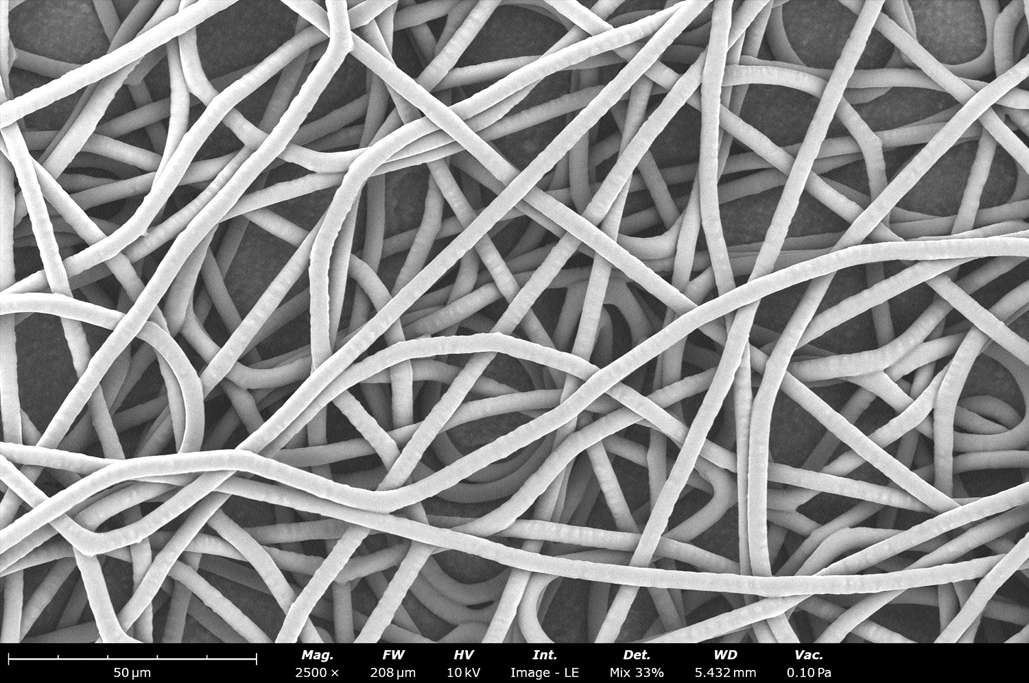
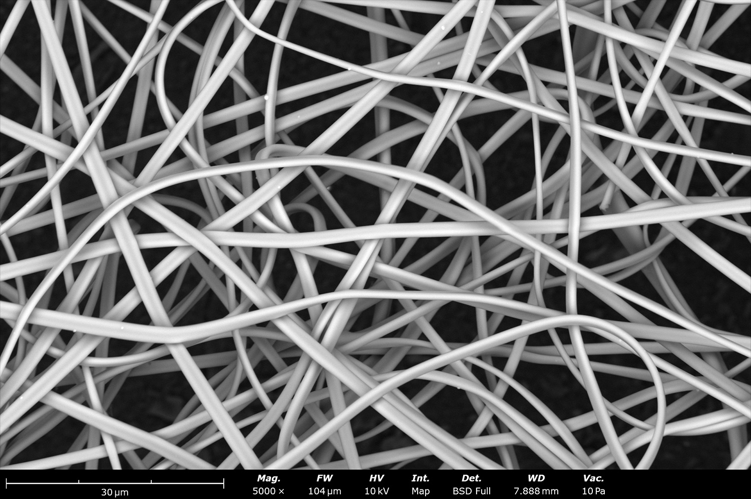
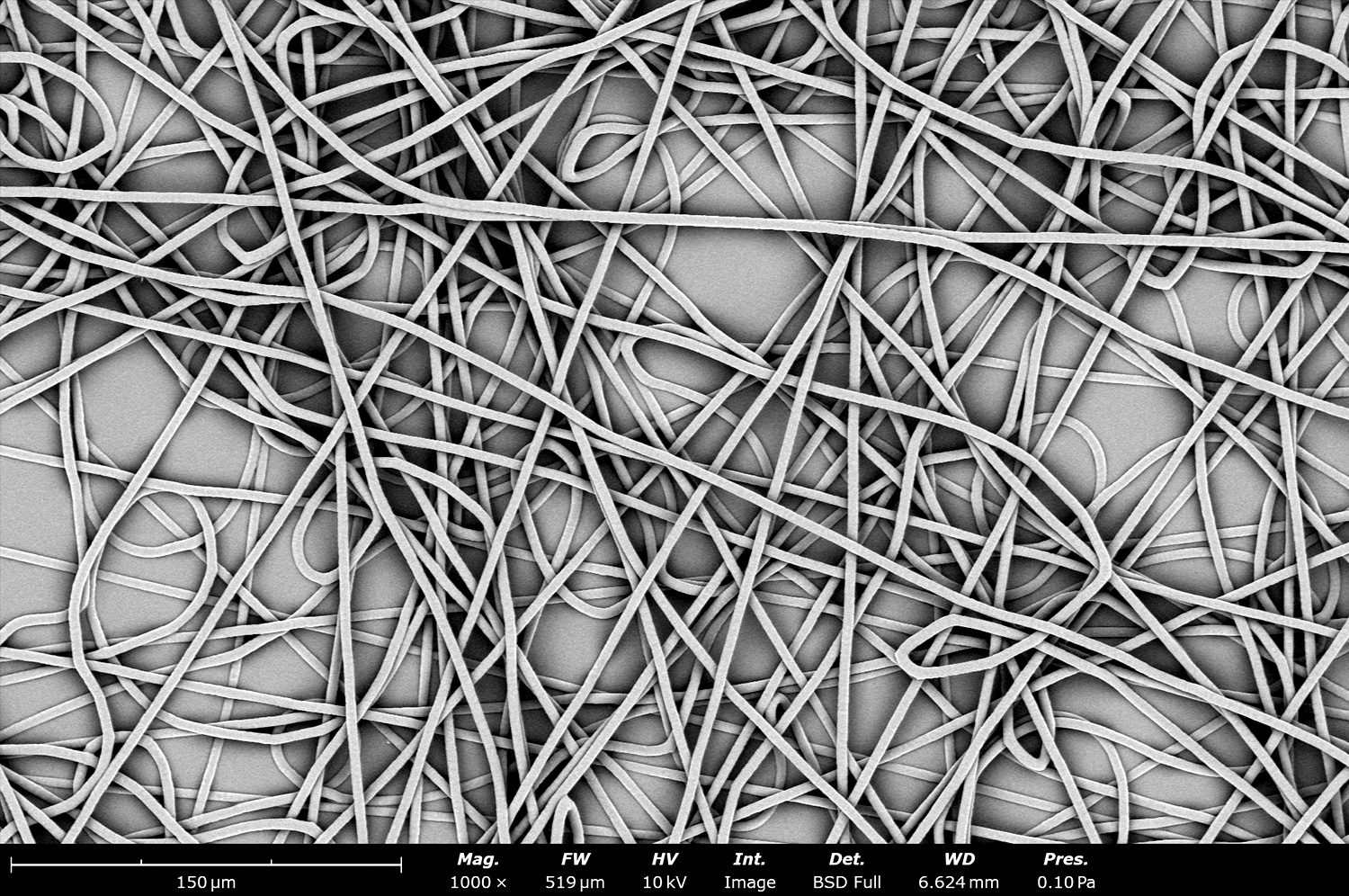
Randomly oriented fibers made out of polycaprolactone (PCL) and on top of a 10 cm diameter drum in a Spinbox equipment. These fibers were collected at 0 rpm to study the effect of fiber orientation under the influence of only the rotational speed in the drum collector.
Randomly oriented fibers made out of polycaprolactone (PCL) and on top of a 10 cm diameter drum in a Spinbox equipment. These fibers were collected at 50 rpm to study the effect of fiber orientation under the influence of only the rotational speed in the drum collector. At this speed the fibers are still randomly oriented throughout the sample structure.
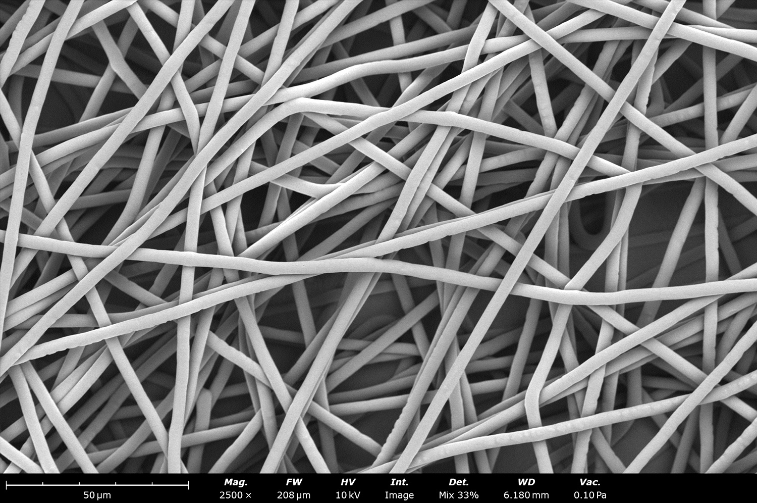
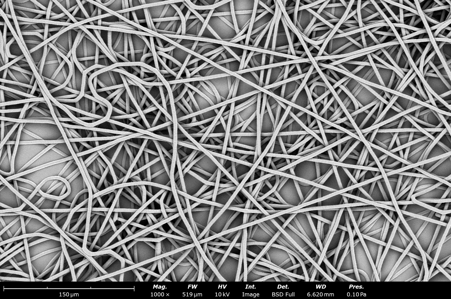
Randomly oriented fibers made out of polycaprolactone (PCL) and on top of a 10 cm diameter drum in a Spinbox equipment. These fibers were collected at 200 rpm to study the effect of fiber orientation under the influence of only the rotational speed in the drum collector. At 200 rpm the PCL fibers are still randomly oriented throughout the sample structure.
Randomly oriented fibers made out of polycaprolactone (PCL) and on top of a 10 cm diameter drum in a Spinbox equipment. These fibers were collected at 500 rpm to study the effect of fiber orientation under the influence of only the rotational speed in the drum collector. At 500 rpm the PCL fibers are still randomly oriented, but some initial reorientation can be seen throughout its microstructure.
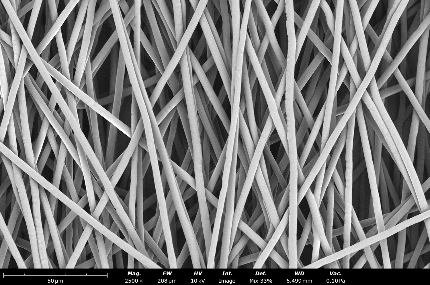
Randomly oriented fibers made out of polycaprolactone (PCL) and on top of a 10 cm diameter drum in a Spinbox equipment. These fibers were collected at 1,000 rpm to study the effect of fiber orientation under the influence of only the rotational speed in the drum collector. At 1,000 rpm the PCL fibers realign during collection and the pore size of the sample is slightly increasing at the same time.
Randomly oriented fibers made out of polycaprolactone (PCL) and on top of a 10 cm diameter drum in a Spinbox equipment. These fibers were collected at 1,500 rpm to study the effect of fiber orientation under the influence of only the rotational speed in the drum collector. At 1,500 rpm the PCL fibers are realigning towards 90 degrees.
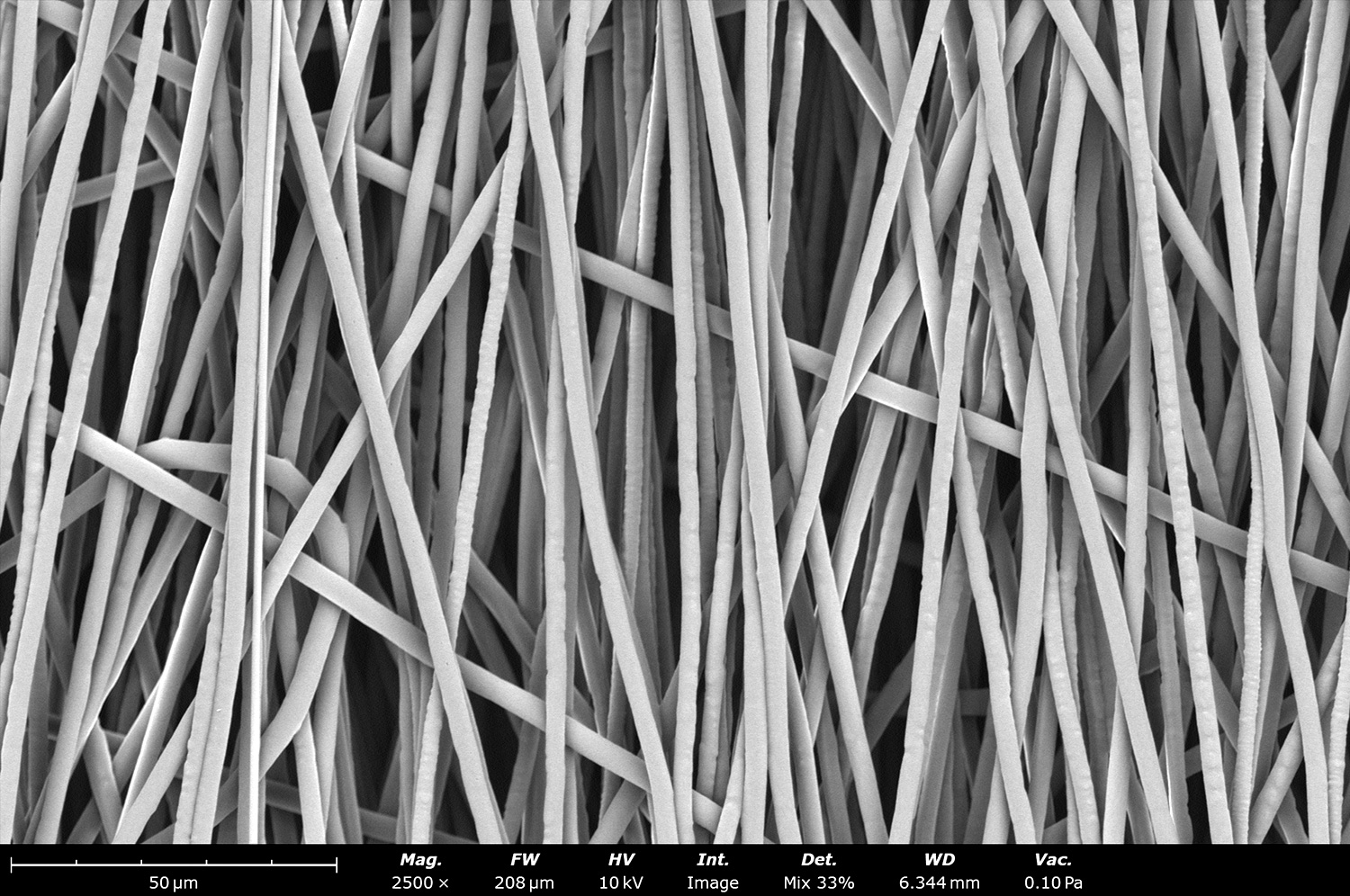
Randomly oriented fibers made out of polycaprolactone (PCL) and on top of a 10 cm diameter drum in a Spinbox equipment. These fibers were collected at 2,000 rpm to study the effect of fiber orientation under the influence of only the rotational speed in the drum collector. At 2,000 rpm the PCL fibers are aligned towards 90 degrees and the fiber diameter has been slightly decreased due to the high linear speed during collection. This orientation is used to increase mechanical properties and biomimic native extracellular matrix of different types of tissue.
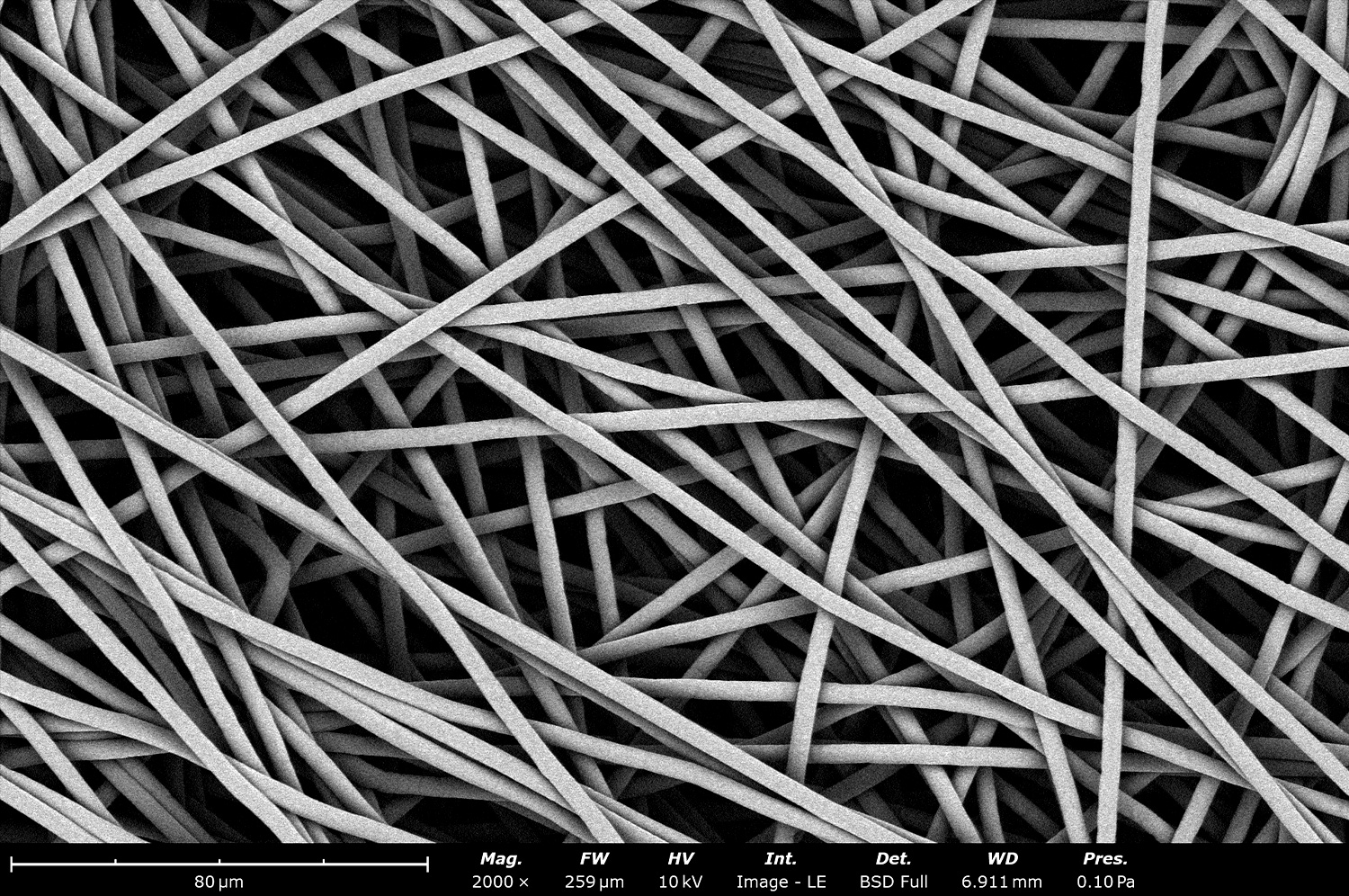
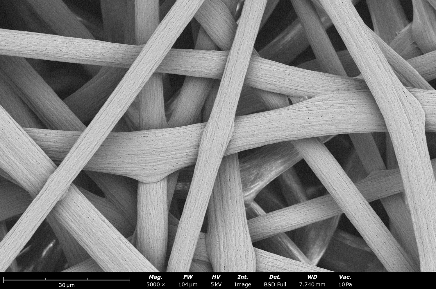
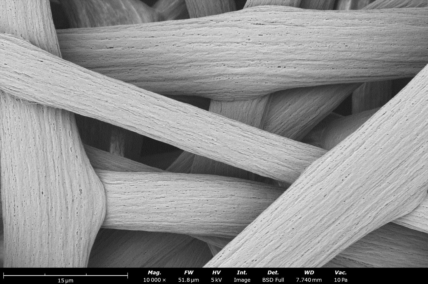
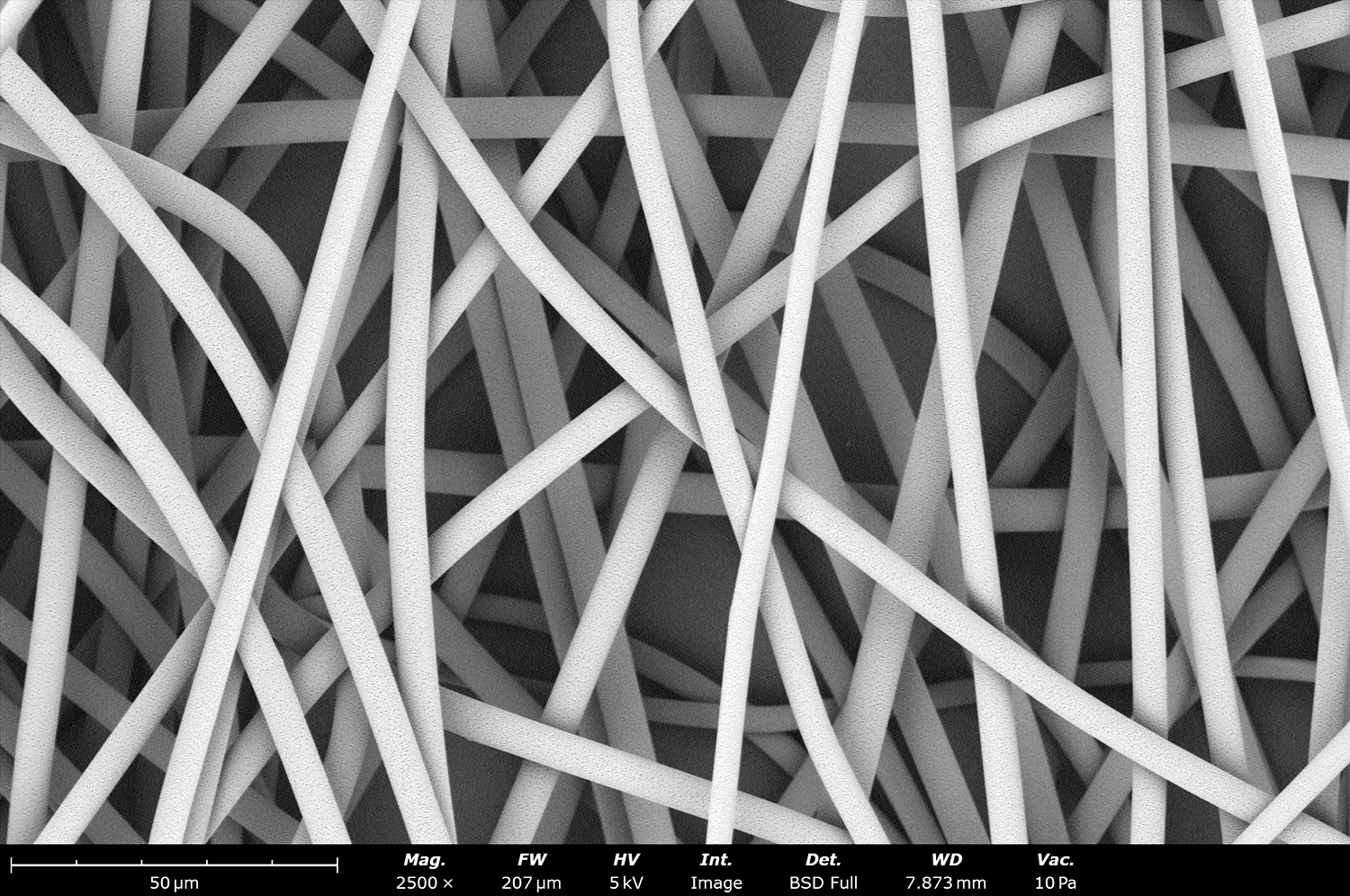
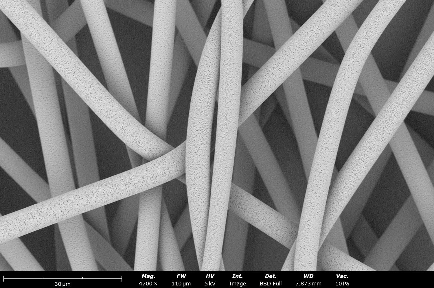
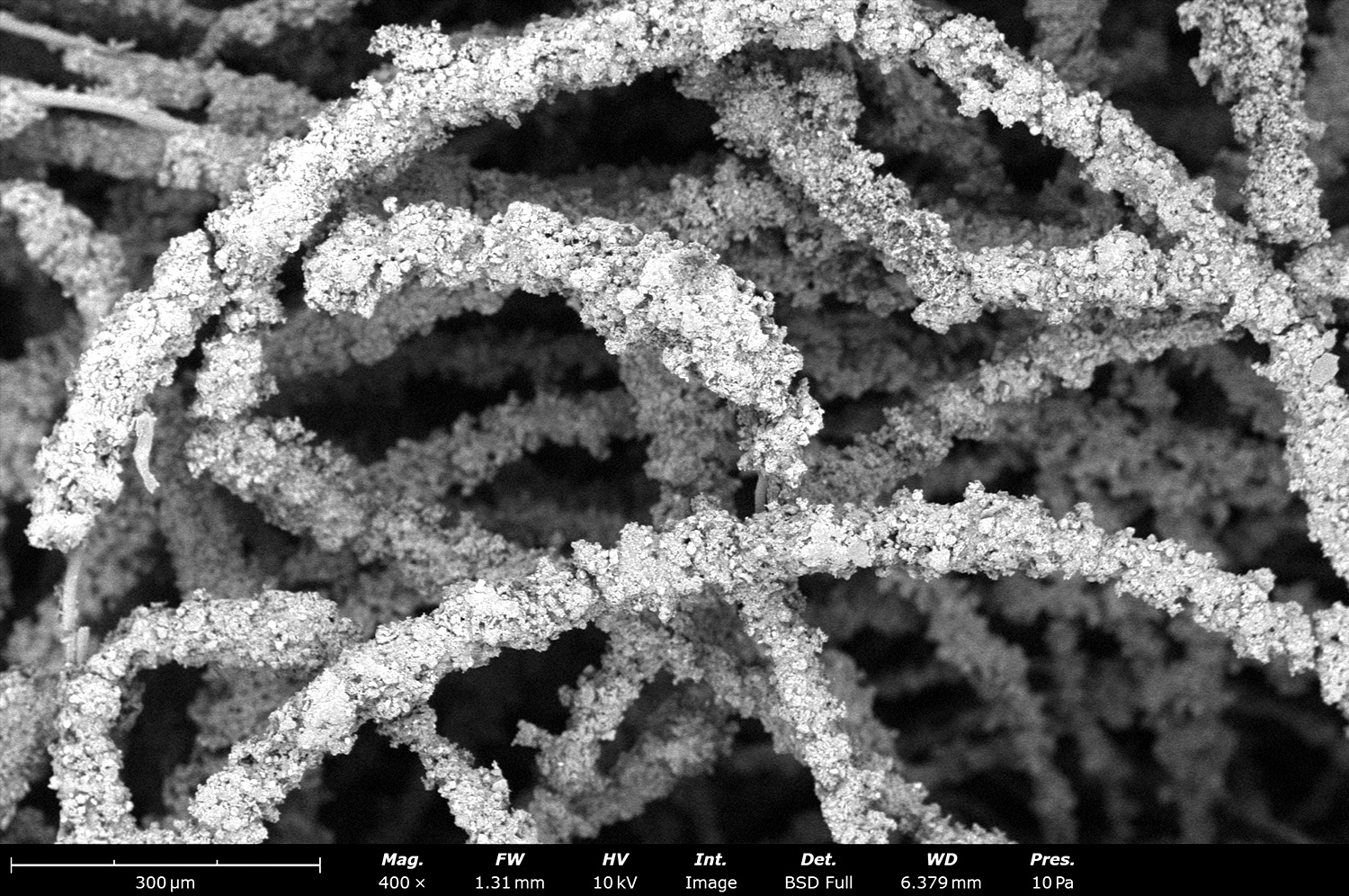
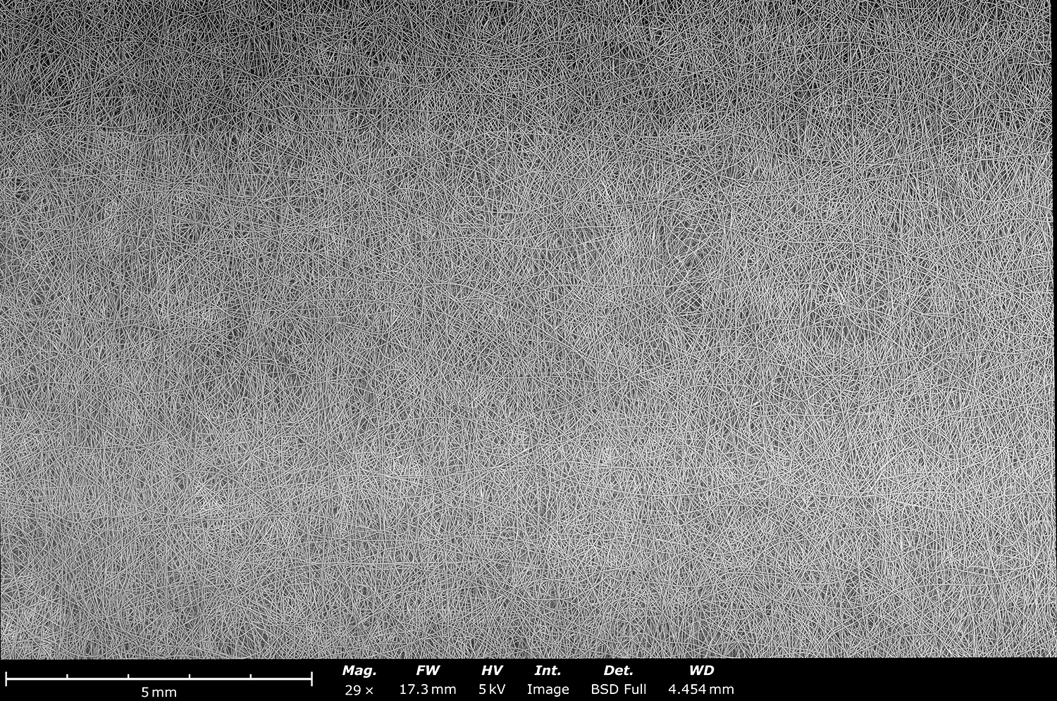
Scanning electron microscope image showing the microstructure of electrospun fiberglass made with a Fluidnatek LE-50. Electrospun fiberglass is being used as a flame retardant material in different applications including textiles. These fibers could also be used as heat dissipation to protect sensitive materials from fire hazards.
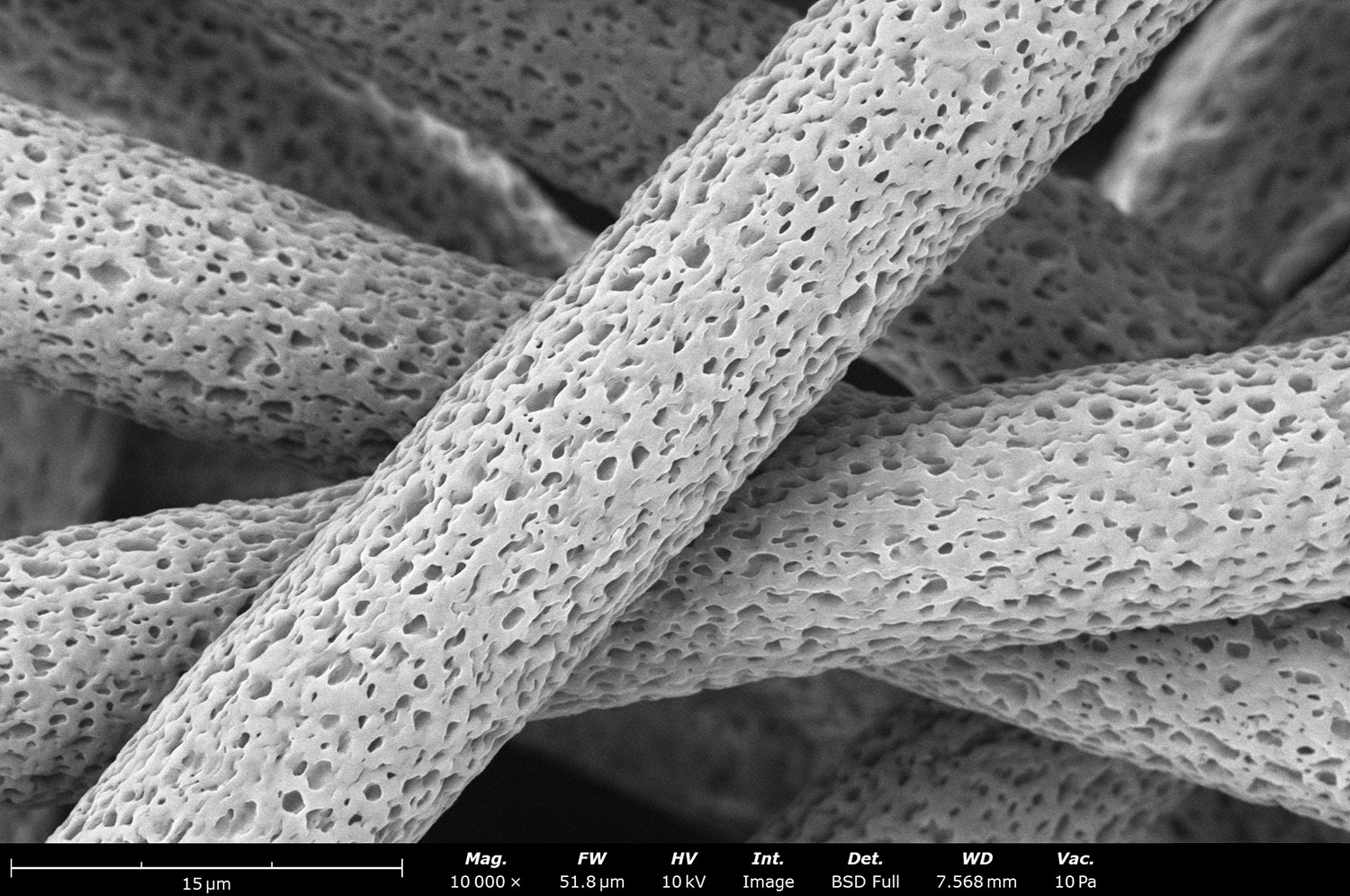
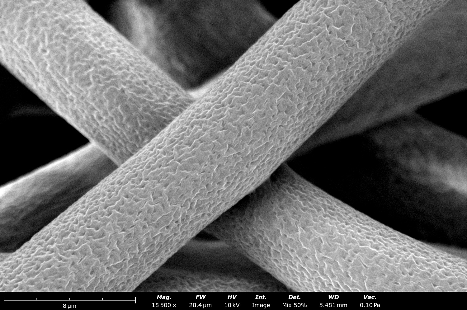
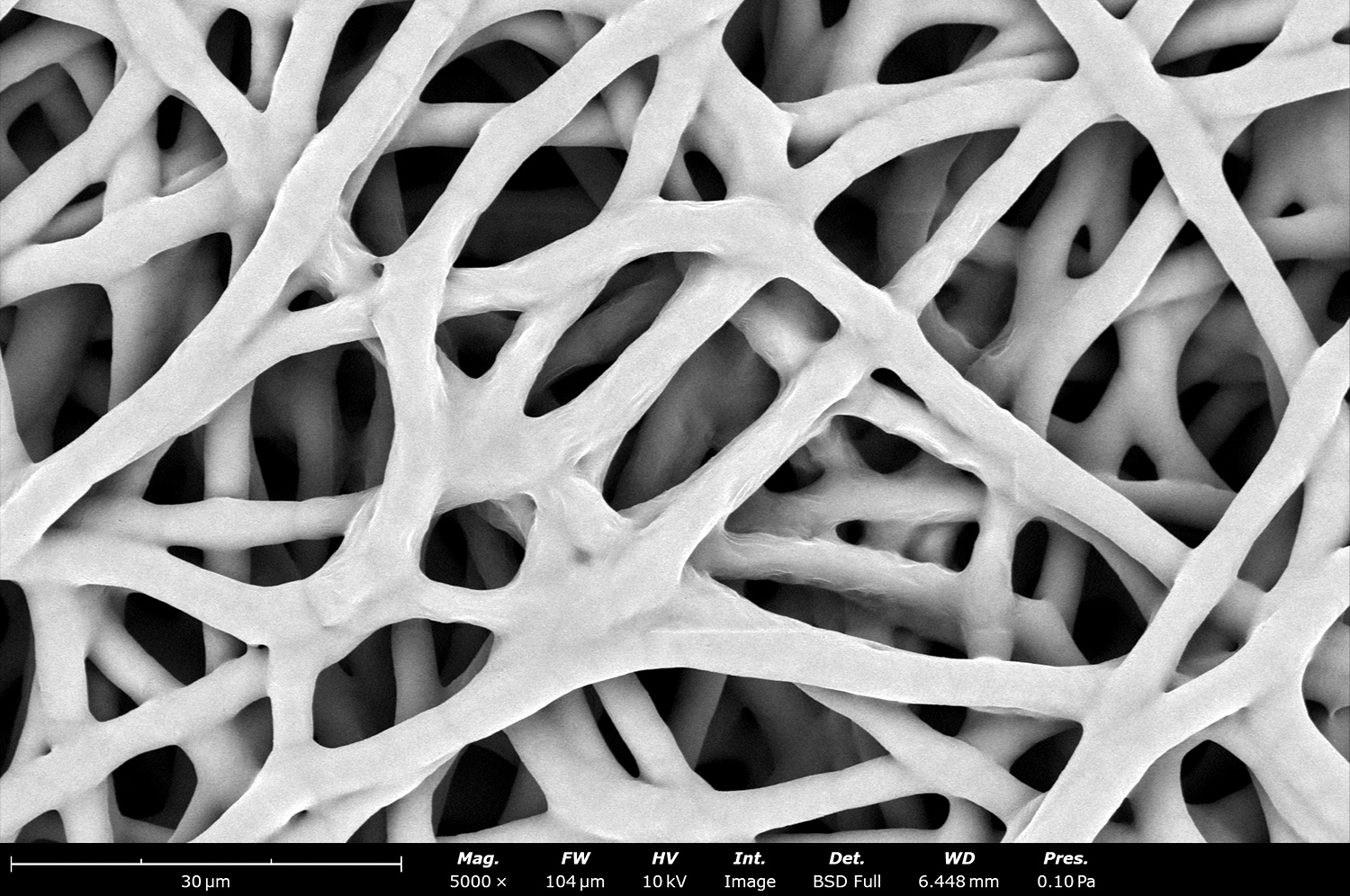
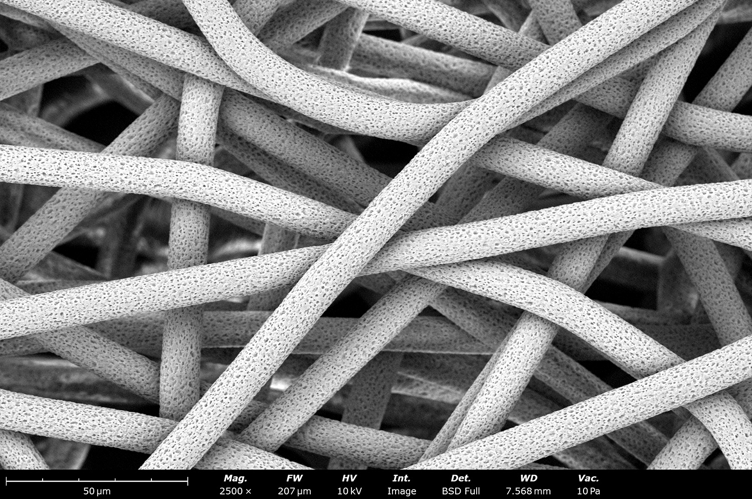
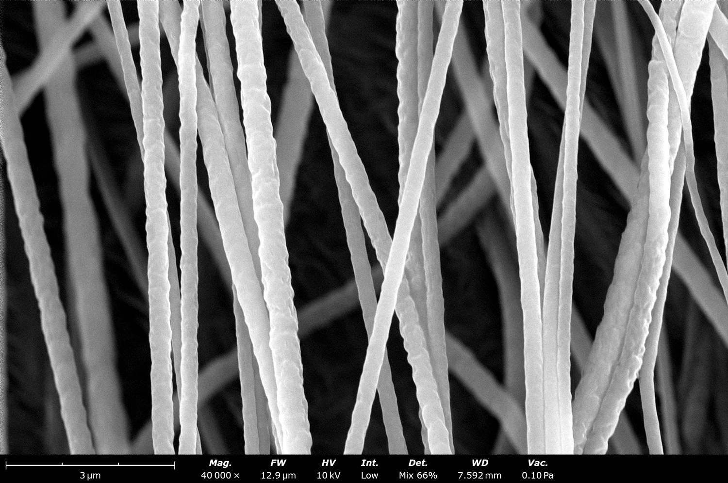
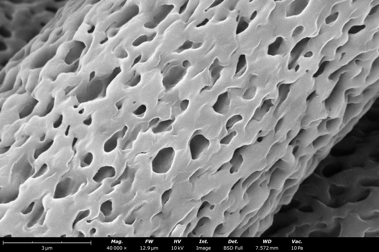
Electron micrograph of biodegradable porous polycaprolactone (PCL) with a diameter around 10 µm. This image shows a close up onto the porous structure of the electrospun PCL fibers. The nanoscale porosity was achieved thanks to the quick solvent evaporation during the electrospinning process.
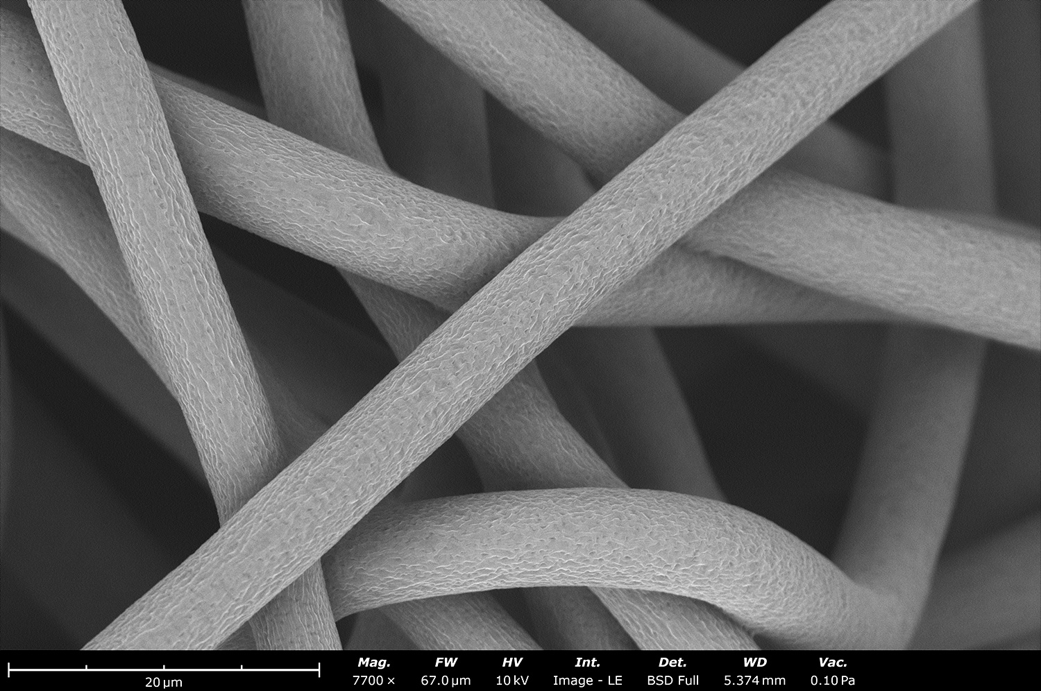
Microstructure of porous polycaprolactone (PCL) fibers with a diameter around 10 µm. These fibers were produced in a single step with the Fluidnatek LE-50 using a single solvent. These large fibers have good elongation properties, making it an ideal material to be used for artificial blood vessels, especially to mimic the tunica media layer of the saphenous vein. The nanoscale porosity was achieved thanks to the quick solvent evaporation during the electrospinning process.
This micrograph shows the cross-section of electrospun polylactide acid (PLA) made using a Spinbox equipment at 1,000 rpm. Thanks to the high glass transition temperature of PLA, the sample preparation to visualize the cross-section only required a simple cut with a scalpel. Other polymers need to be freeze fractured to prevent smearing from happening. Electrospun PLA fibers are typically used for wound healing and drug delivery applications, along with coating medical devices.
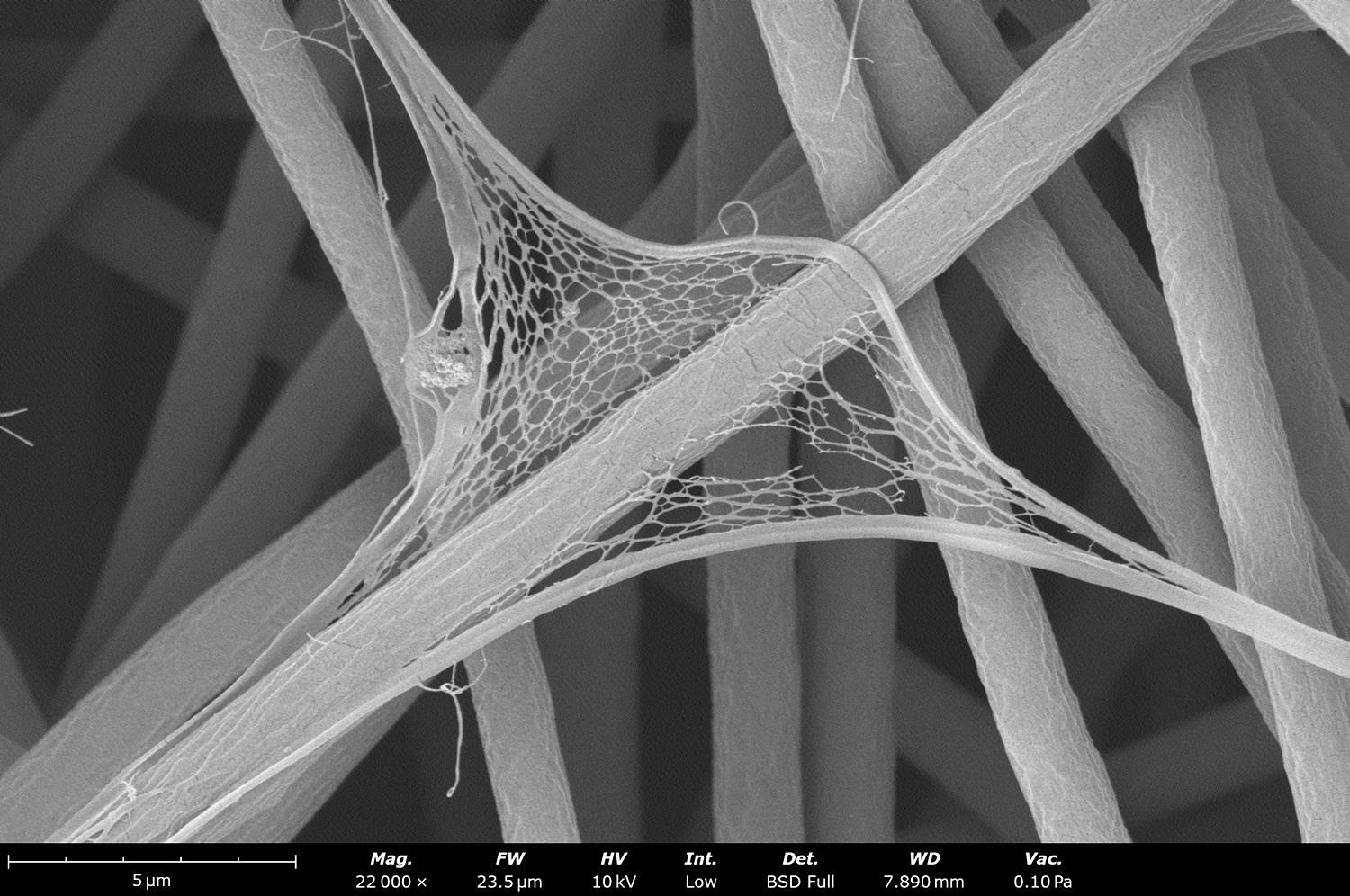
SEM image of electrospun polycaprolactone (PCL) made using the Fluidnatek LE-50. This image was acquired with a backscatter electron detector at an accelerating voltage of 5 kV. The microstructure reveals fiber-fiber bonding. This is due to low collector voltage during sample processing. When fibers are attracted faster to the collector, the solvent has less time to evaporate, allowing fibers to be wet and generate fiber-fiber bonding.
Coronary artery stent with an outside diameter of 5 mm coated with electrospun polycaprolactone (PCL). This SEM image is a composite of 5 rows and 8 columns stitched automatically to have a representative look at the microstructure of the coated surface. Stents are coated with fibers to make them more biocompatible. The beauty of the electrospinning technique is that it allows stents to be coated with different polymeric materials and drugs can be incorporated to improve patient healing
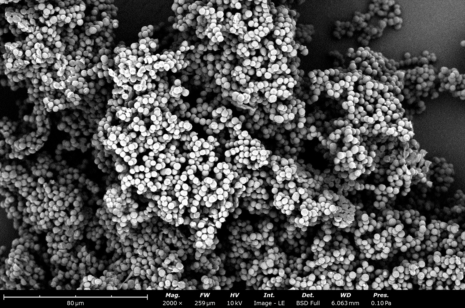
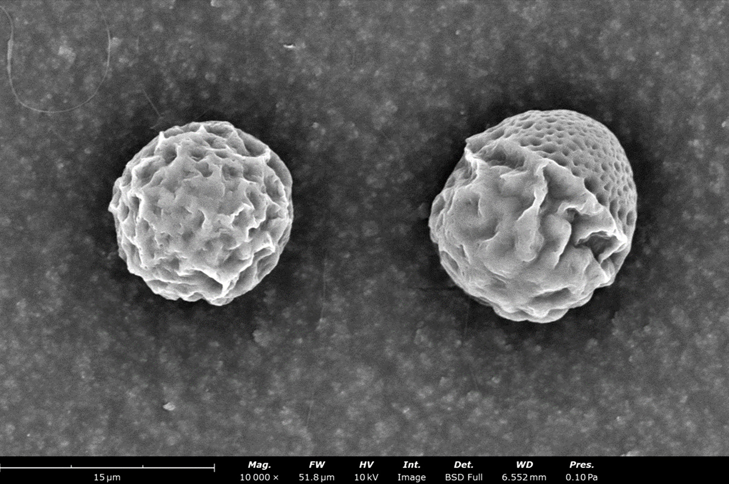
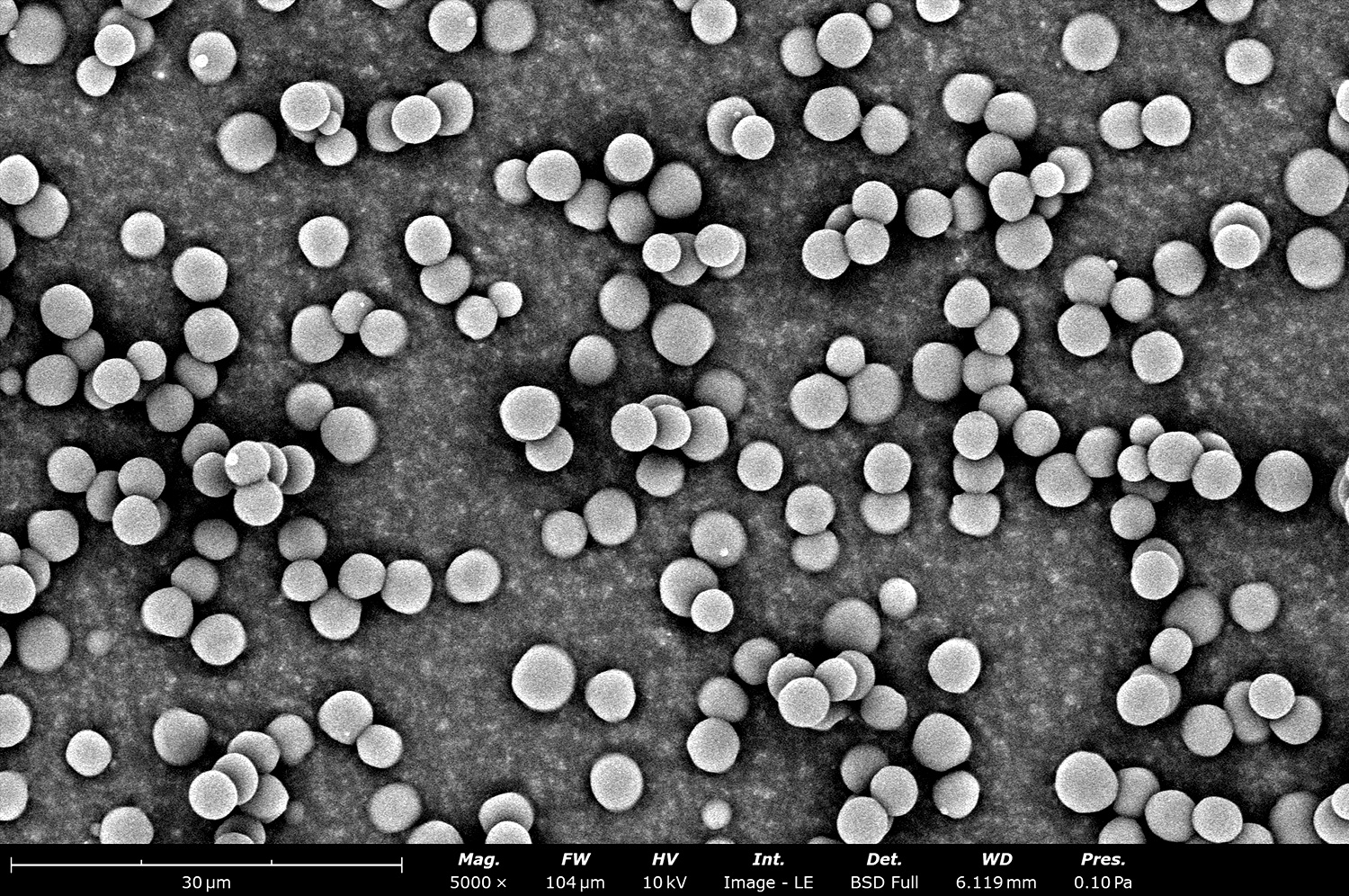
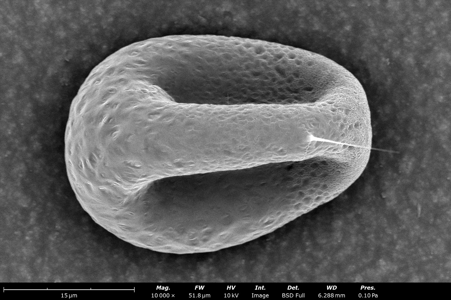
Backscattered electron micrograph of electrosprayed polycaprolactone (PCL) particles made with a Fluidnatek LE-50 and collected onto a liquid reservoir. These electrosprayed particles are characterized by their collapsed structure and the nanoscale porosity, both due to the use of a low boiling point and high vapor pressure solvent, along with their large diameter (greater than 20 µm). These PCL particles can be used for drug delivery applications and to uniformly coat medical devices
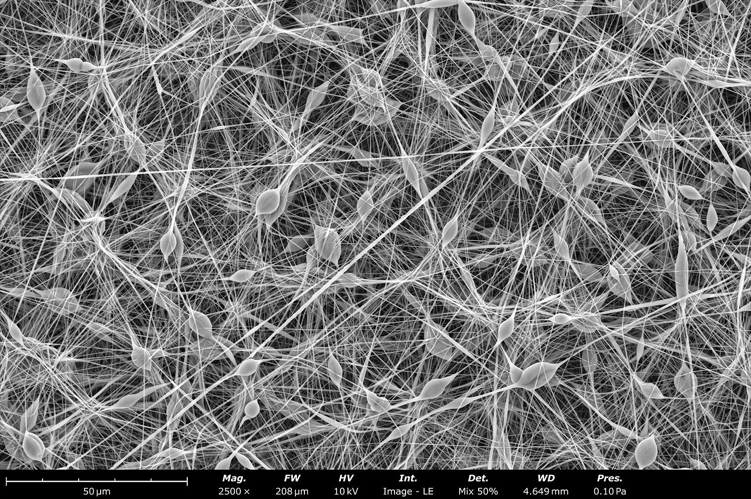
These electrosprayed polycaprolactone (PCL) rough microparticles were made on a single step using a Spinbox equipment and collected onto a flat plate collector covered with aluminum foil. Note the two distinctive surfaces (Janus morphology) on the right particle. Increasing the surface area of a particle, especially without post-processing and at room temperature, can help with drug delivery applications by loading thermal sensitive materials
Stitched image of 40 scanning electron micrographs of polycaprolactone (PCL) fibers collected on top of a 1 cm diameter rod at 200 rpm and with the collector grounded in a Fluidnatek LE-100 equipment. At 0 kV on the collector a rough microstructural surface is observed due to residual charge, causing fibers to localize in certain areas of the sample, affecting the final porosity of the electrospun material. By using a higher bias this effect will be completely removed and the final sample will contain a smooth surface.
Backscattered electron micrograph of polycaprolactone (PCL) fibers collected on top of a 1 cm diameter rod at 200 rpm in a Fluidnatek LE-100 equipment. At 200 rpm, and for large deposition times, a rough microstructural surface is observed that is due to residual charge, causing fibers to localize in certain areas of the sample, affecting the final porosity of the electrospun material. This microstructural effect is removed when using higher linear speeds or when combined with a higher bias at the same rotational speed.
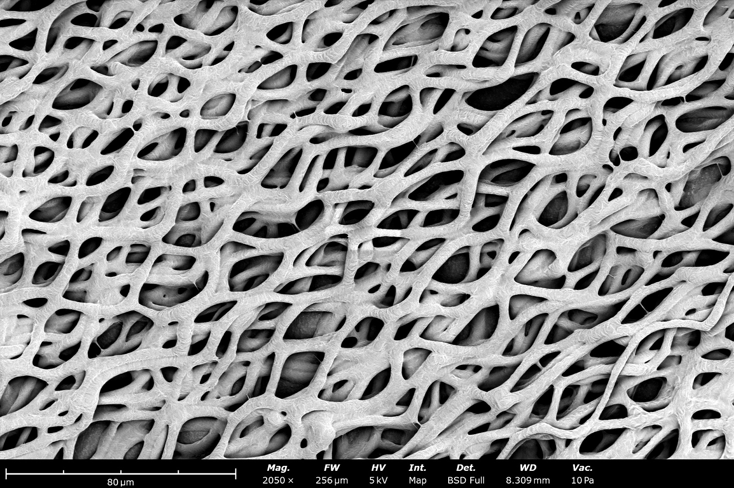
This SEM image shows the microstructure of ribbon-shaped gelatin fibers made out of electrospinning with a Fluidnatek LE-50 and collected onto a rotating drum. The ribbon-shape structure is typically observed when the solvent evaporates fast during the spinning process. The fast evaporation process causes the fibers to collapse and obtain this flattened structure. A rounded morphology can be obtained by optimizing solution properties by using a high boiling point solvent like acetic acid.
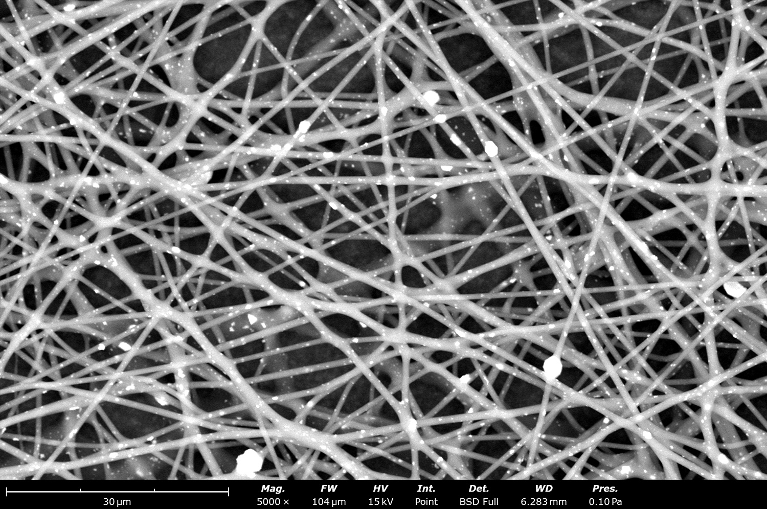
Backscattered electron micrograph of electrospun nylon 6,6 (N66) fibers made with a Fluidnatek LE-500 with a multi-needle system and continuously collected onto a roll-to-roll collector. N66 is a commonly used polyester for air and liquid filtration applications due to its hydrophilicity, good solvent resistance, tailored fiber diameter, pore size tuning and ease of scalability with the electrospinning technology.
Micrograph of electrospun Nylon nanofibers made with a Fluidnatek LE-100 and collected onto a flat plate collector. Nylon nanofibers are commonly used for filtration applications, tissue engineering like bone regeneration when incorporated with hydroxyapatite, and medical sutures as they are stronger and durable when compared with silk.
The effect of fiber deposition density and microstructure was studied using electrospun polycaprolactone (PCL) fibers, collecting onto a flat plate collector of a Fluidnatek LE-500 by having all processing parameters constant and only changing the collector voltage. With a grounded collector (0 kV) we can see a low fiber density and large pore size.
The effect of fiber deposition density and microstructure was studied using electrospun polycaprolactone (PCL) fibers, collecting onto a flat plate collector of a Fluidnatek LE-500 by having all processing parameters constant and only changing the collector voltage. With a -1 kV applied in the collector, we can see a denser deposition when compared to the 0 kV sample. All fibers look relatively dry at these processing conditions.
The effect of fiber deposition density and microstructure was studied using electrospun polycaprolactone (PCL) fibers, collecting onto a flat plate collector of a Fluidnatek LE-500 by having all processing parameters constant and only changing the collector voltage. With a -5 kV applied in the collector, we can see a denser deposition when compared to the -1 kV sample. All fibers look relatively dry at these processing conditions.
The effect of fiber deposition density and microstructure was studied using electrospun polycaprolactone (PCL) fibers, collecting onto a flat plate collector of a Fluidnatek LE-500 by having all processing parameters constant and only changing the collector voltage. With a -10 kV applied in the collector, we can see that the fibers are not fully dried as there is more fiber-fiber contact, and the pore size has been reduced.
Backscattered electron micrograph of electrospun polycaprolactone (PCL) fibers, collecting onto a flat plate collector of a Fluidnatek LE-500 with a collector voltage of -10 kV. Fiber-fiber bonding is clearly seen throughout the microstructure. Although typically considered a non-desired defect, these wet fibers can be used to adhere two layers of materials with the electrospinning technique. Dry fibers can be obtained after adhesion by simply changing the collector voltage towards 0 kV.
Electron micrograph of electrosprayed polycaprolactone (PCL) microparticles made with a Fluidnatek LE-50. These particles have a diameter around 4 µm and were collected on top of polyethylene with carbon black to allow ease of removal post-processing. This smooth surface can be made with other types of polymers to obtain dry particles in a single step, encapsulate materials, and increase throughput by scaling up the electrospraying process for industrial production.
This SEM image shows an electrosprayed polycaprolactone (PCL) microparticles made with a Fluidnatek LE-50 using a low boiling point and high vapor pressure solvent. These particles show a characteristic collapsed surface with pores throughout its diameter ranging from a couple microns to the nanoscale size.
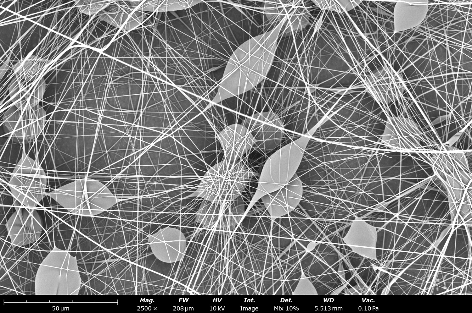
Microstructure of a sample made by combining electrospun polycaprolactone (PCL) nanofibers and electrosprayed PCL microparticles using the Fluidnatek LE-100. Applications like tissue engineering can benefit from combining fibers and particles in a single step and at room temperature conditions to have encapsulated growth factors that can be released at specific times. These types of samples can also be used for drug delivery applications on medical devices.
This SEM image shows the microstructure of polycaprolactone (PCL) nanofibers when combined with PCL electrosprayed particles at the same time in the Fluidnatek LE-100. To achieve this, the sample was collected onto a rotating drum by electrospinning the fibers in a horizontal setup and electrospraying the particles in a vertical setup, both needles pointing towards the same collector.
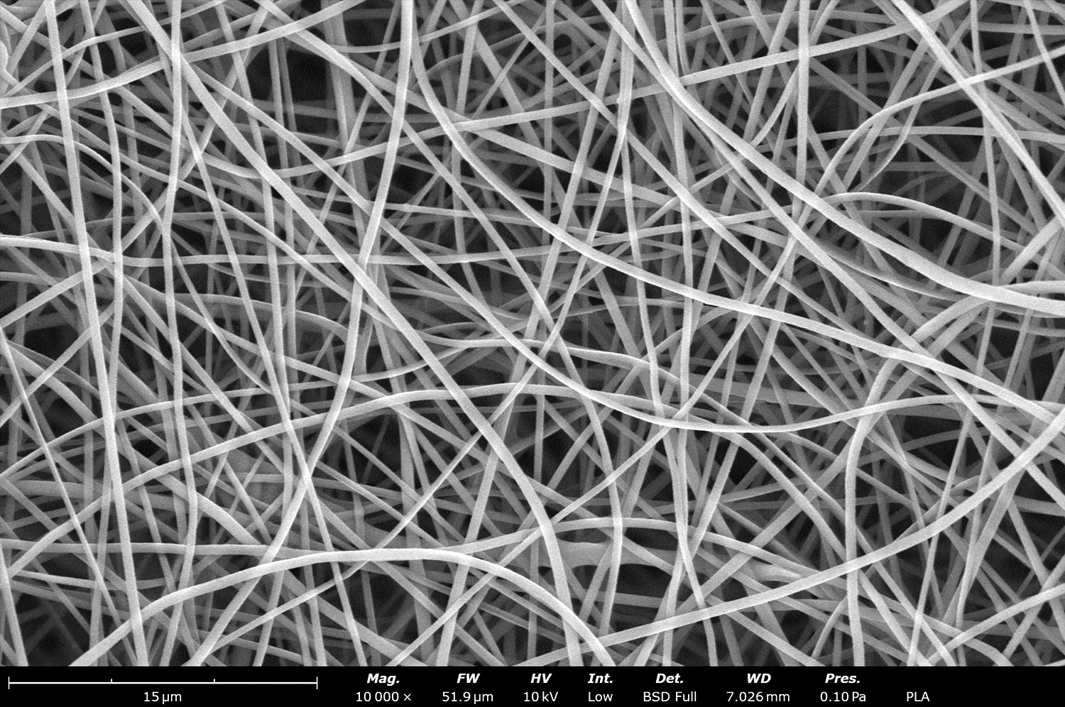
Microstructure of electrospun polylactic acid (PLA) fibers with sub-micron diameter. These fibers were produced in a single step with the Fluidnatek LE-50 using a solvent mixture of dichloromethane and dimethyl formamide solvent. PLA is used in FDA approved medical products, making it an ideal material to be used for artificial blood vessels, covering stent or for wound healing processes.
Electrospun collagen fibers using 1,1,1,3,3,3-hexafluoroisopropanol (HFIP) as the solvent. Collagen is commonly used in wound healing applications as it is a polymer found in the in vivo environment. This sample was developed using the Fluidnatek LE-50 and under tight environmental conditions (23°C and 60% relative humidity) to allow for ease of processability.
Fiberglass is typically use in applications were durability and heat resistance is a must. These electrospun beaded fibers were made with the Fluidnatek LE-50 using the rotating collector platform to generate a sample of 20 cm x 32 cm.
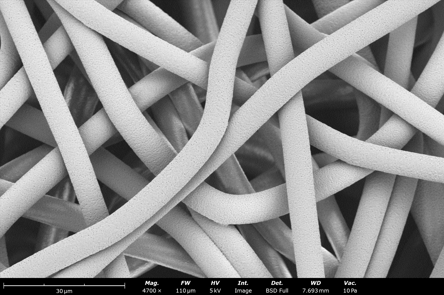
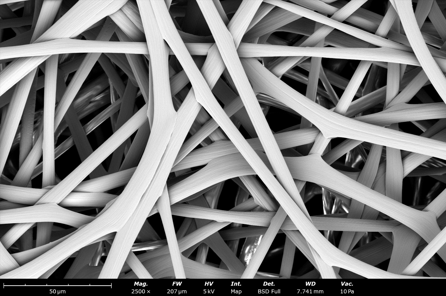
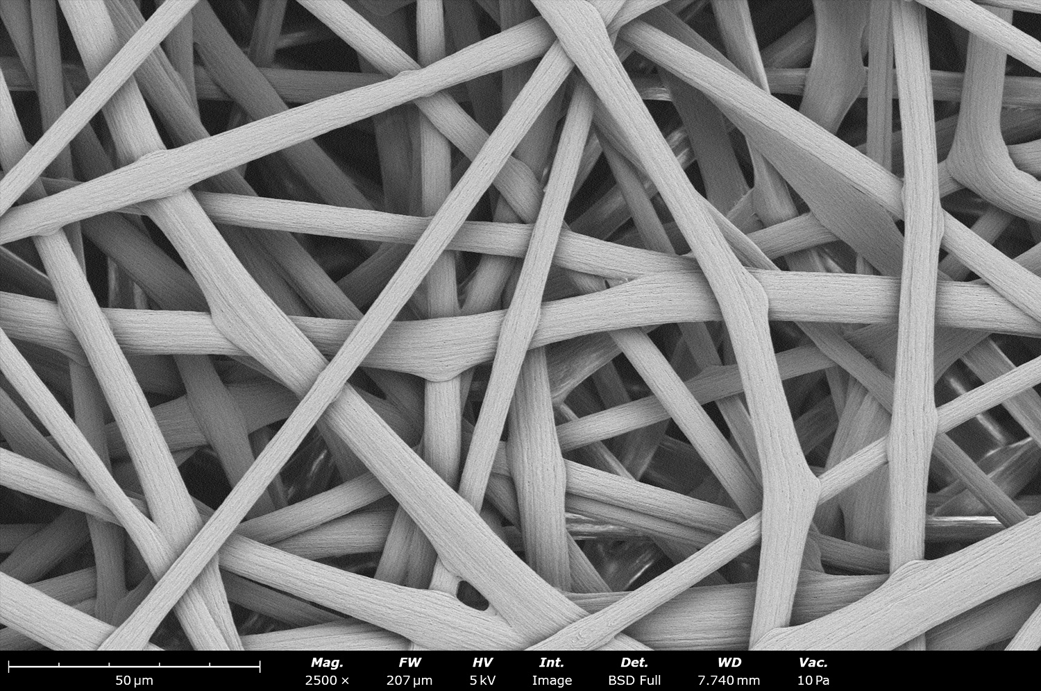
Microstructure of polycaprolactone (PCL) microfibers with a diameter around 8 µm. These fibers were produced with dichloromethane (DCM). Since DCM has a low boiling point and high vapor pressure, it evaporates fast during sample processing, generating large fibers. The final sample has large pore size, allowing the sample to be an ideal candidate for tissue engineering applications as cells will be able to infiltrate and proliferate in the material
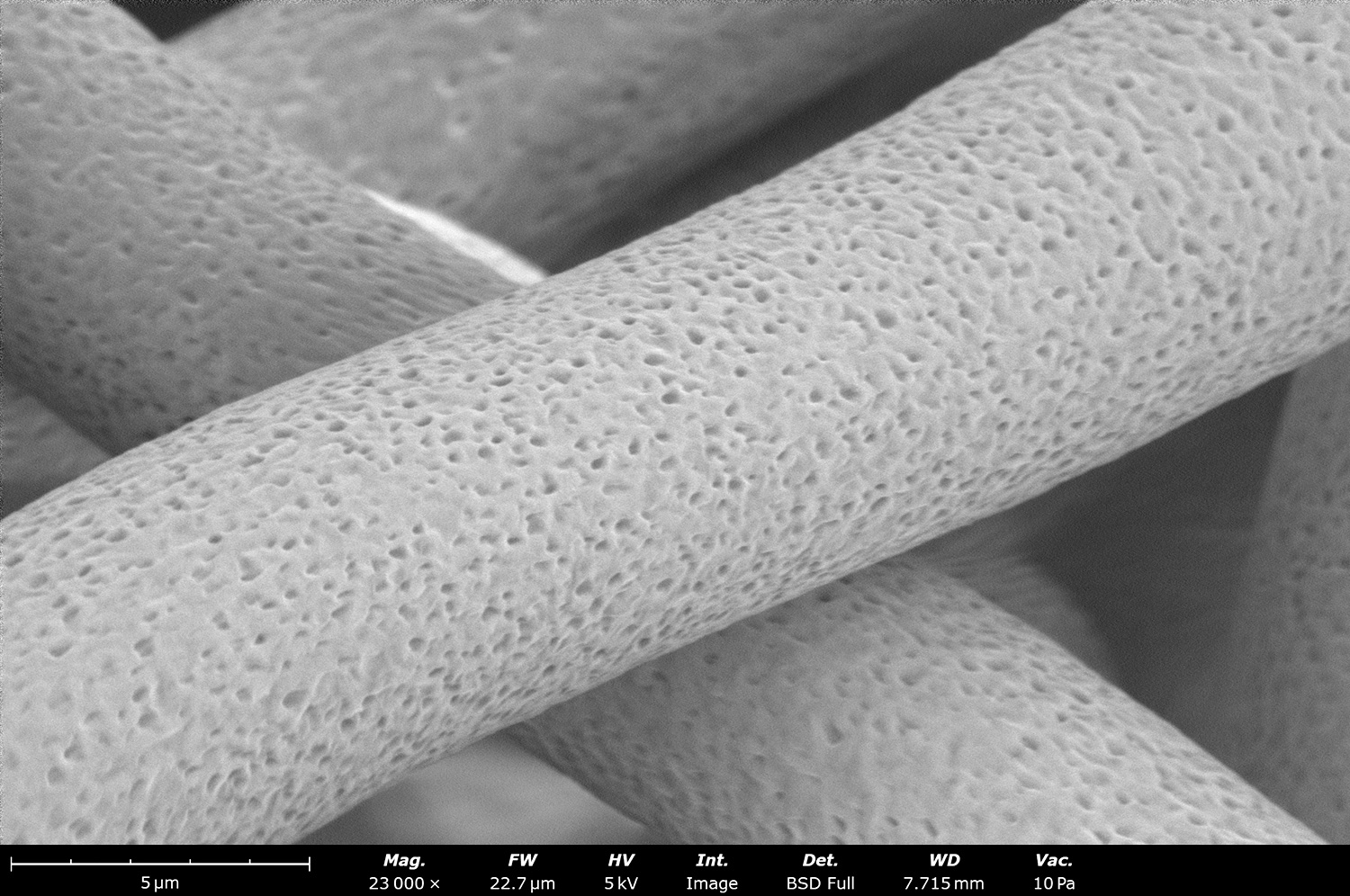
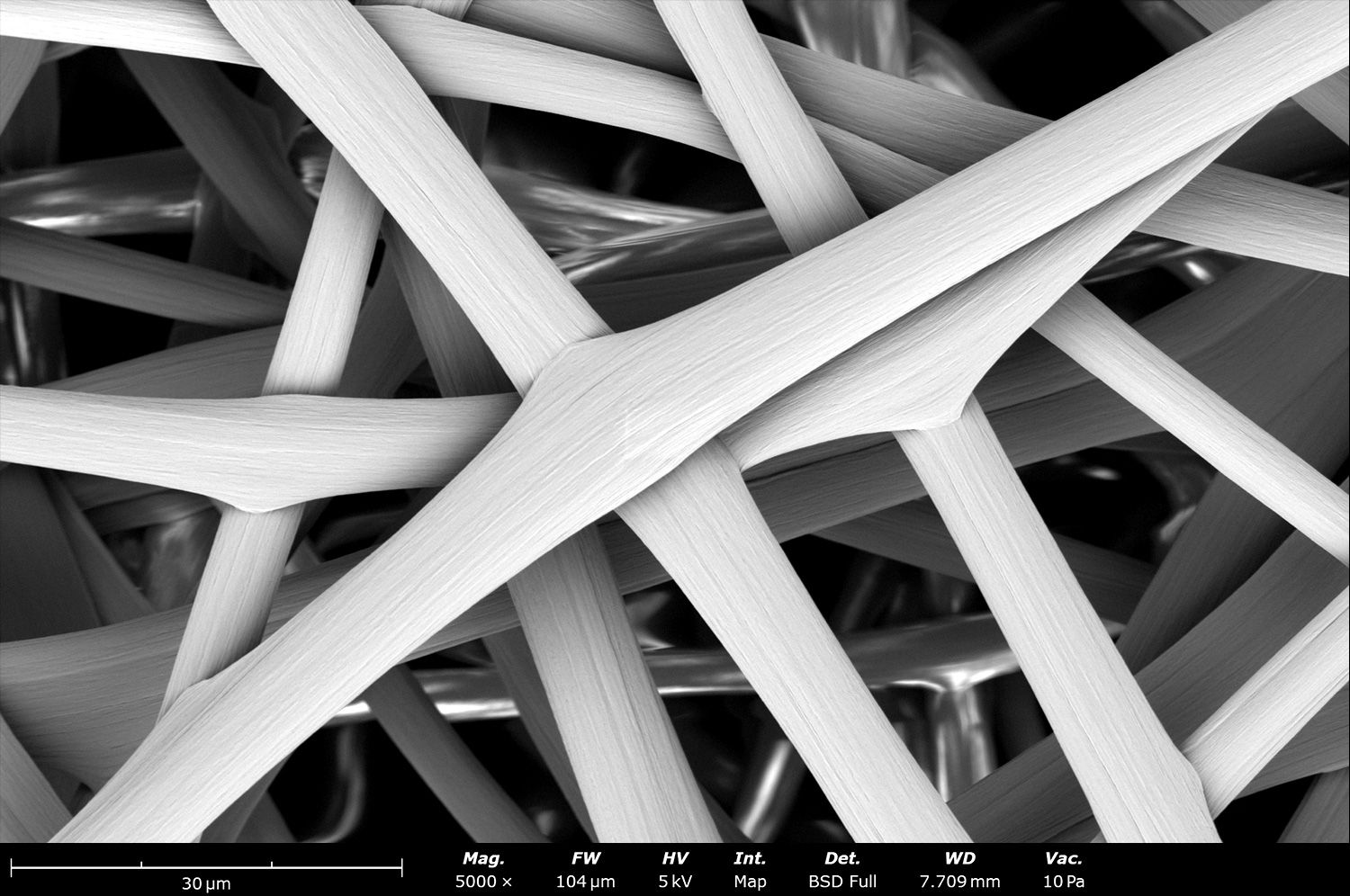
Upon close inspection, we see a porous structure on these 8 µm polycaprolactone (PCL) fibers. The small porosity allows cells to anchor and proliferate easier. These fibers were made using the Fluidnatek LE-50 using a rotating drum collector of 10 cm in diameter at 200 rpm.
Electrospinning allows the use of blending materials during sample processing. This image shows electrospun microscale fibers composed of polycaprolactone (PCL) and Nylon 6,6 (N66). Before processing, these two polymers were blended with the same solvent. During processing the final material is a combination of PCL:N66 that will impart properties to fulfill different application needs.
This microstructure shows microscale fibers of blended polycaprolactone (PCL) and polyethylene terephthalate (PET). The combination of PCL and PET is typically used when mechanical properties on the final structure are needed.
This SEM image taken on the Phenom Desktop SEM shows the microstructure of blended polycaprolactone and polyethylene terephthalate microfibers. These PCL:PET fibers are also combined when the degradation rate of PCL needs to be fine-tuned by adding a biostable PET, or when PET fibers need to be in combination with a biodegradable polymer like PCL
Post-processing as-spun electrospun fibers is typically done when microstructure or properties of the final sample are needed. This image shows the microstructure changes of PCL:PET fibers when treated with acetone for 5 seconds. At a magnification of 2,500 X the microstructure looks similar to the as-spun PCL:PET fibers.
Upon close inspection we can see smaller pores on the surface of the fibers when the sample is imaged at 5,000 X. This porosity could change microstructure properties, drug release properties, or even cell proliferation depending on application needs
Microstructure of acetone post-treated microfibers of PCL:PET. Imaging at 10,000 X we can clearly see the nanoscale porosity that has appeared on the surface of the electrospun fibers. Depending on the polymer compatibility, blended ratio and interaction during processing, different type of post-processing structures could be obtained and fine tuned based on application needs.
Microstructure of microscale polylactic acid (PLA) fibers with an average diameter of 6 µm. These fibers were produced in a single step with the Fluidnatek LE-50 using a single solvent. These large fibers have good elongation properties, making it an ideal material to be used for artificial blood vessels, especially to mimic the tunica media layer of the saphenous vein.
Electron micrograph of biodegradable porous polylactic acid (PLA) with an average diameter of 6 µm made with dichloromethane (DCM) as the solvent in solution. This image reveals a porous structure of the surface of the electrospun PLA fibers. The nanoscale porosity was achieved thanks to the quick solvent evaporation during the electrospinning process.
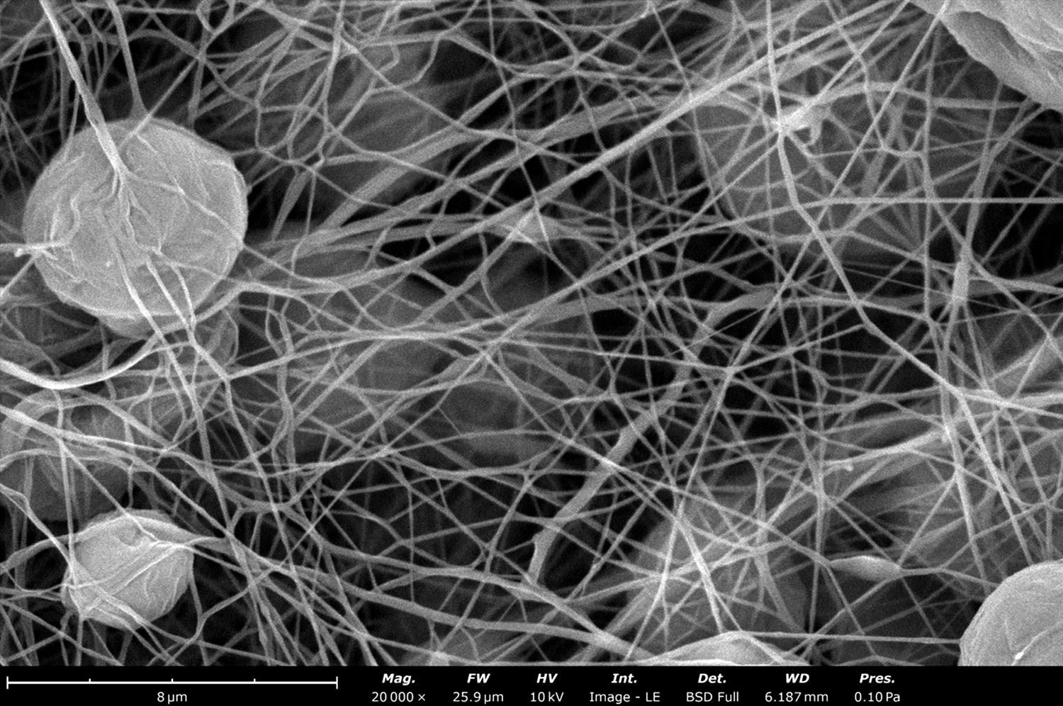
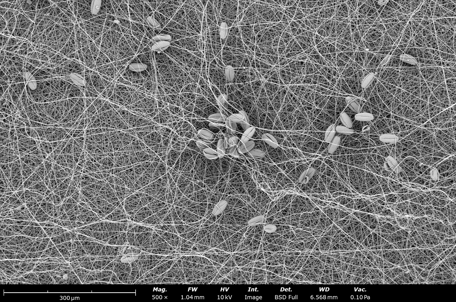
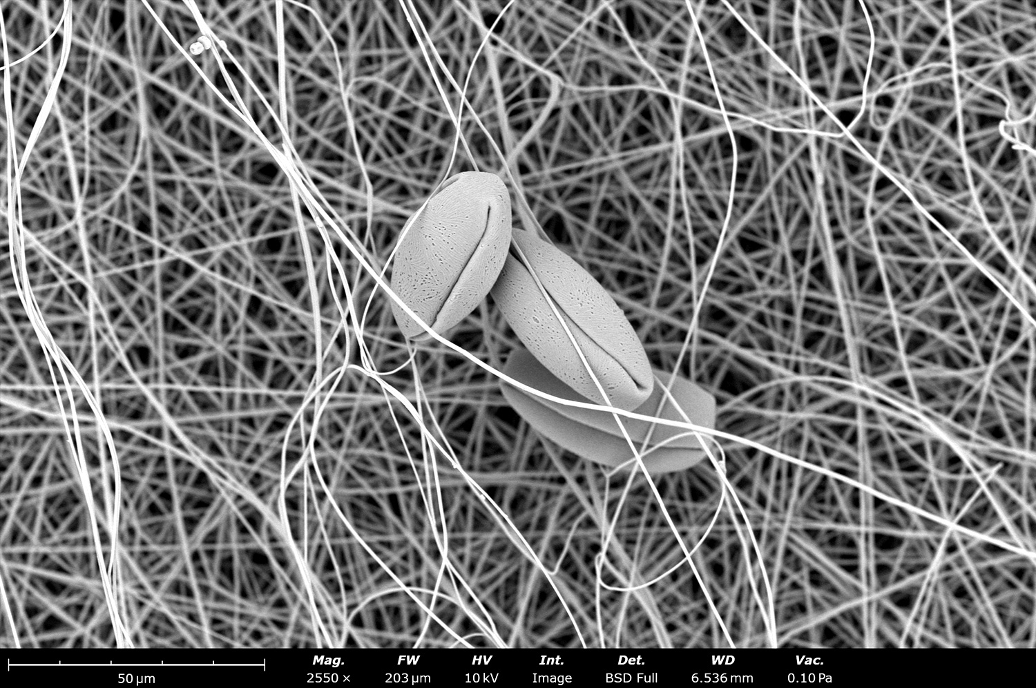
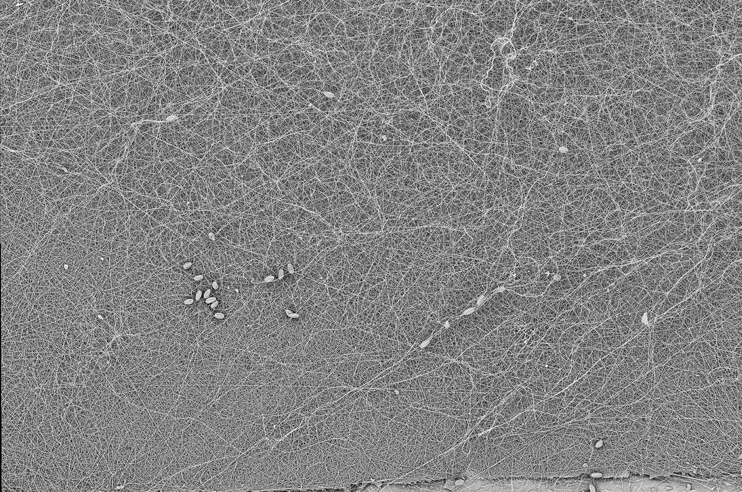
Microstructure of Nylon 6,6 (N66) nanoscale fibers with trapped pollen on its surface. Nylon 6,6 is commonly used for filtration applications. Electrospinning allows the generation of nanoscale N66 fibers allowing for easy mechanical entrapment of particles on filtration media.
Electrospun micrograph showing trapped pollen on the nanoscale fibers of Nylon 6,6 (N66) made with electrospinning. N66 is an easy to process polymer through the electrospinning technique. It has been used in scale up processes to generate more than 1,000 m2/h of material.
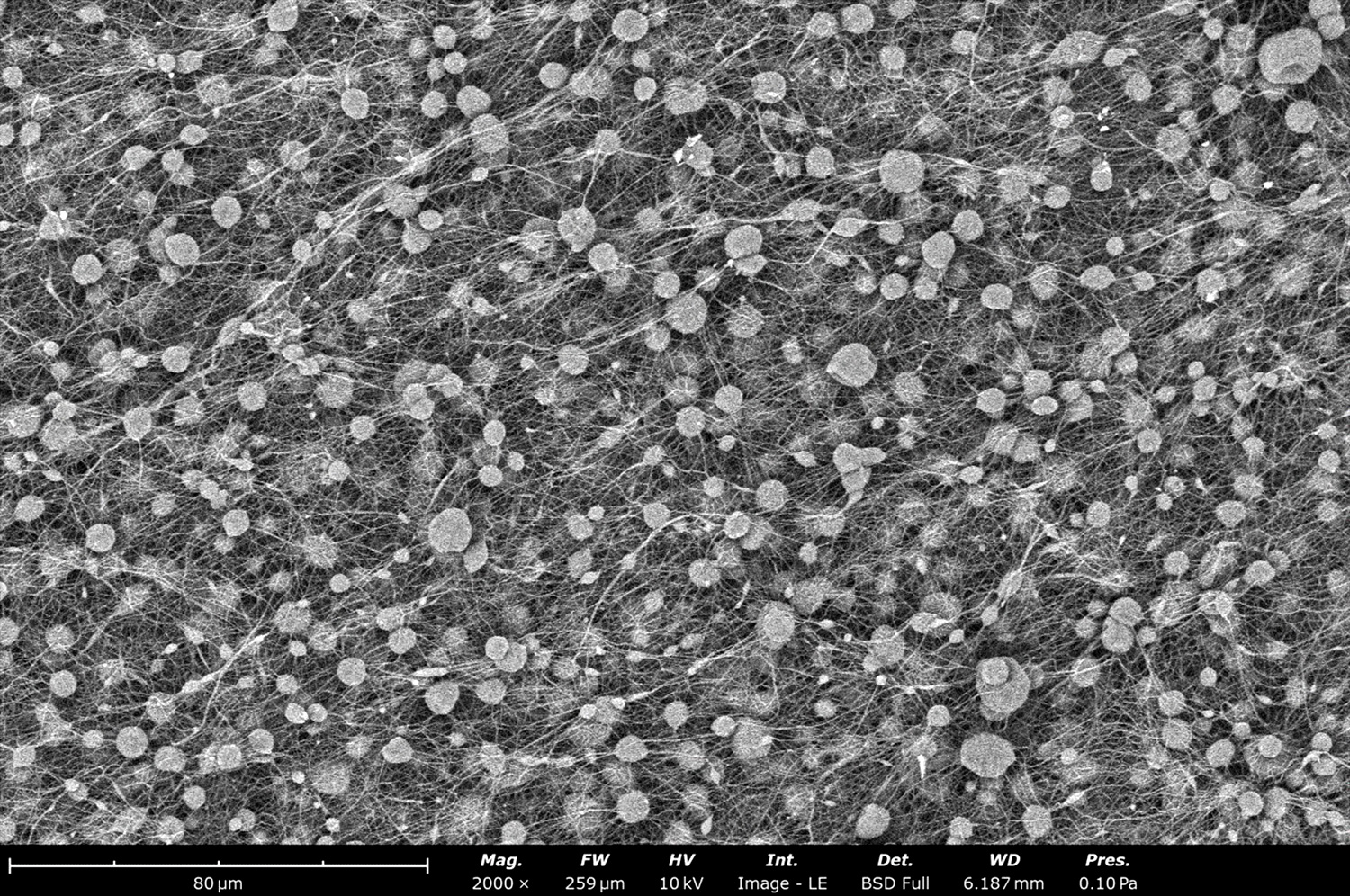
This image shows an overall look on the filtration media made out of Nylon 6,6 with trapped pollen on its structure. Electrospun fibers allow for mechanical filtration and samples that can be reused when compared to statical filtration media.
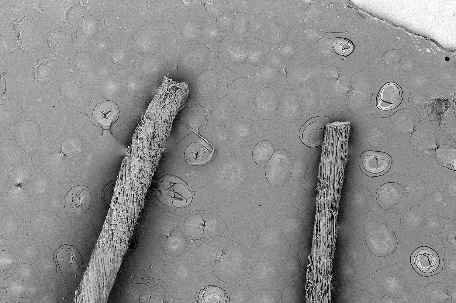
The microstructure of thermoplastic polyurethane (TPU) fibers generated with the Fluidnatek LE-50 using a mandrel collector of 5 mm in diameter. TPU fibers are commonly used in medical application as it is a flexible polymer with good mechanical properties. The electrospinning technique allows for fine tuning the fiber diameter and pore size to fulfill different application needs.
Electron micrograph of electrospun thermoplastic polyurethane (TPU) fibers. This image shows a combination of micron and sub-micron scale TPU fibers made with the electrospinning technique. TPU is a polymer used in medical product currently approved and cleared by the Food and Drug Administration (FDA).
Overall view of a surgical mesh coated with electrospun thermoplastic polyurethane (TPU) fibers. TPU fibers were used to coat the surface of the surgical mesh to make it more biocompatible by allowing the structure to resemble the extracellular matrix. By using electrospinning, the addition of fibers can make the surgical mesh more biocompatible and accepted by the body after implantation.
This stitched SEM image shows the microstructure of TPU fibers when electrospun on top of a surgical mesh for a long time. The ability of electrospinning to coat different substrates with different thickness give the flexibility to obtain desired properties based on application needs.
This image shows the back area of the surgical mesh coated with electrospun TPU fibers. Electrospinning has the ability to properly attach electrospun fibers to any type of mesh and prevent delamination. This allows the final sample to not go over post-processing needed with typical techniques used by the scientific community.
Microstructure image of electrospun hyaluronic acid (HA), a natural polymer typically used in food packaging, cosmetics, and tissue engineering applications. This image with a magnification of 10,000 X reveals sub-micron fibers. The analysis was performed with a mixture of backscattered and secondary detector at a ratio of 50:50.
This image shows the microstructure of electrospun nanoscale polyethylene oxide (PEO) fibers. These fibers were developed with the Fluidnatek LE-50 using a rotating drum of 10 cm in diameter and rotating at 100 rpm. The solvent used for processing PEO was water. To prevent needle clogging or dryness, the environmental conditions were maintained at 28°C and 40% relative humidity.
Electron micrograph of electrospun polyethylene terephthalate (PET) beaded fibers. Beaded fibers are usually not desired by the scientific community. Meanwhile, beaded fibers offer advantages like encapsulation ability of different active ingredients or drugs that can be delivered without bursting release. In order to prevent beaded fibers in the final morphology, the easy solution is to increase the concentration of the final solution. This will allow you to obtain only fiber formation without beads.
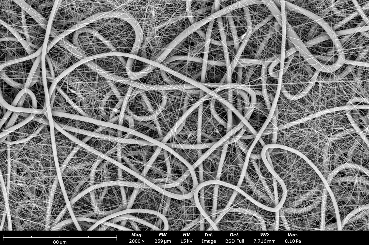
This SEM image shows the microstructure of nanoscale electrospun fibers out of polyvinylidene fluoride (PVDF). Nanoscale fibers are commonly developed with the electrospinning technique, and we can develop them with a diameter less than 20 nm depending on solution properties. These fibers were made with the Fluidnatek LE-50 using the rotating drum of 10 cm in diameter and at 50 rpm to obtain randomized morphology.
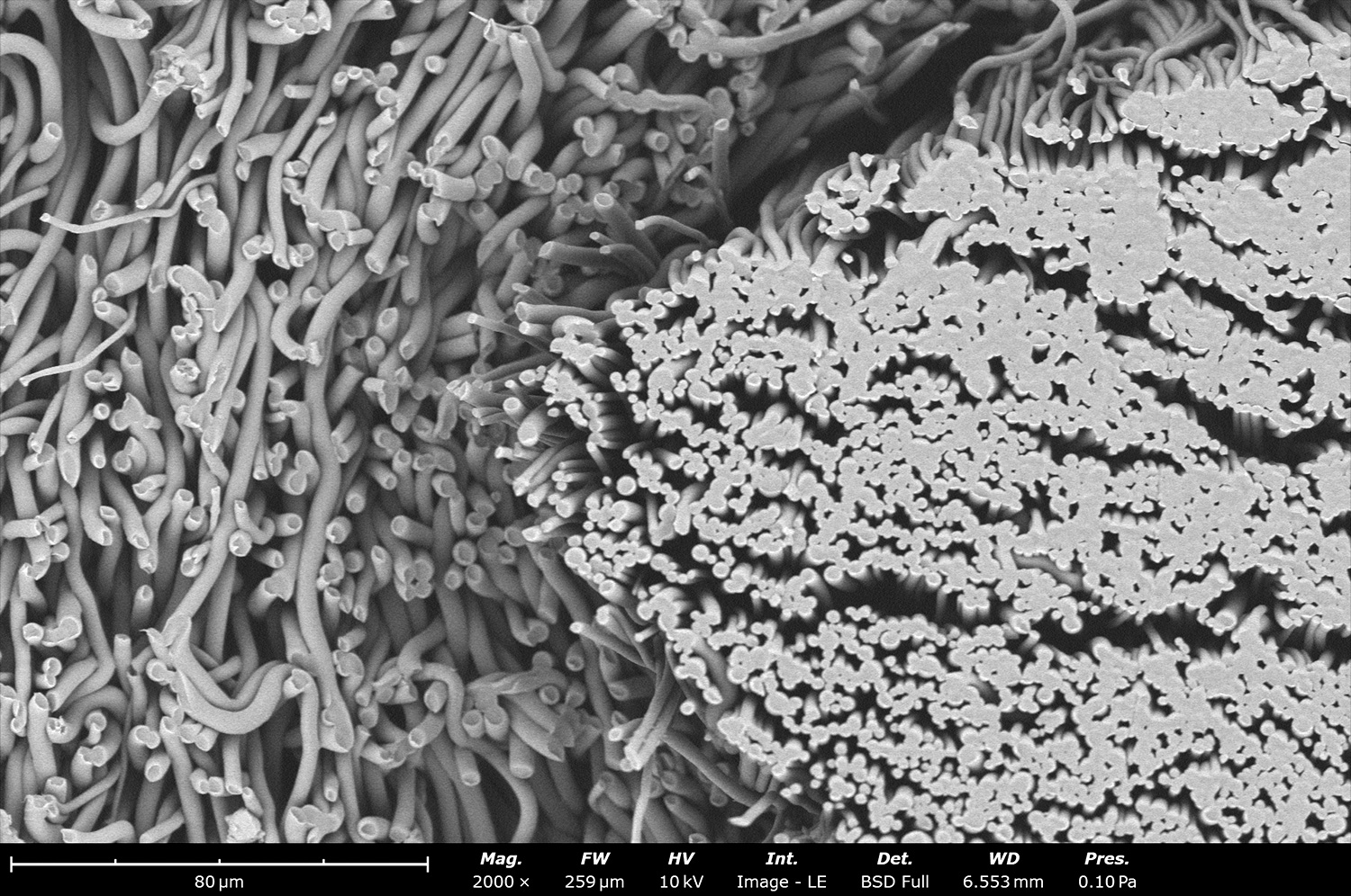
Microstructure of a non-woven material covered with electrosprayed particles using the Fluidnatek LE-500. Electrospraying can be used to coat different types of materials and surfaces to impart different properties that the native material does not have by itself. As seen in the SEM image, the particles only cover the non-woven material and not the porosity of the structure, opening many possibilities for the coating industry.
Electron micrograph of a 200 µm diameter metal wire of coated with electrospun fibers made out of a synthetic polymer and using the Fluidnatek LE-100 BioTubing. The Fluidnatek technology allows to cover samples with diameters less than 1 mm with precise thickness and uniformity. Coating different surfaces allows to impart properties like biocompatibility, drug delivery form the fibers, improve mechanical strength improvement, among others.
This image SEM image shows a cross-sectional image of an electrospun nerve graft made out of three distinctive layers of poly(lactic acid) fibers. Electrospinning has the innate capability to biomimic different morphologies that are found on native tissue. Artificial nerve grafts are one of the many possibilities of microstructure that can be made with the Fluidnatek technology and the electrospinning technique.
Backscattered electron micrograph of electrospun polyacrylonitrile (PAN) in DMSO made using the Fluidnatek LE-50. The image shows an intended effect of fast attraction to the collector when using a low negative voltage. A low voltage can help prevent delamination between two layers but is always recommended to expose the sample to a vacuum environment to remove residual solvent.
Microstructure of polyacrylonitrile (PAN) in DMSO using the Fluidnatek LE-50. This image shows the effect of negative voltage when fibers are attracted fast towards the collector. Lower collector high voltage can be used to induce bundles, obtain larger fiber diameter and have residual solvent that will help with adhesion between two layers. Beaded structures are seen as the fiber did not bend and whip enough to stretch the material during collection.
Backscattered electron micrograph of electrospun poly(n-butyl methacrylate) (PBMA) microfibers generated with the Fluidnatek LE-50. PBMA is a biocompatible polymer used to coat metal stents and control drug delivery on those types of devices. The microstructure of PBMA can be easily tuned with electrospinning to generate nano- or micro-fibers, with different pore size and porosity, based on application needs.
This image shows the microstructure of poly(vinylidene fluoride-co-hexafluoropropylene) (PVDF-HFP) electrospun fibers made with the Fluidnatek LE-50. PVDF-HFP is a commonly used thermoplastic polymer in applications like medical device and binding for electrodes in lithium-ion batteries. In medical applications, PVDF-HFP is used to prevent blood clotting and inhibit platelet adhesion.
Overall view of a surgical mesh coated with a thin layer of electrospun polycaprolactone (PCL) microfibers. PCL fibers were used to coat the surface of the surgical mesh to make it more biocompatible as the microstructure resembles the extracellular matrix of native tissue. By using electrospinning, the addition of fibers can make the surgical mesh more biomimetic and accepted by the body after implantation.
This stitched SEM image shows the microstructure of PCL fibers when electrospun on top of a surgical mesh for a long time. The ability of electrospinning to coat different substrates with different thickness gives the flexibility to obtain desired properties based on application needs.
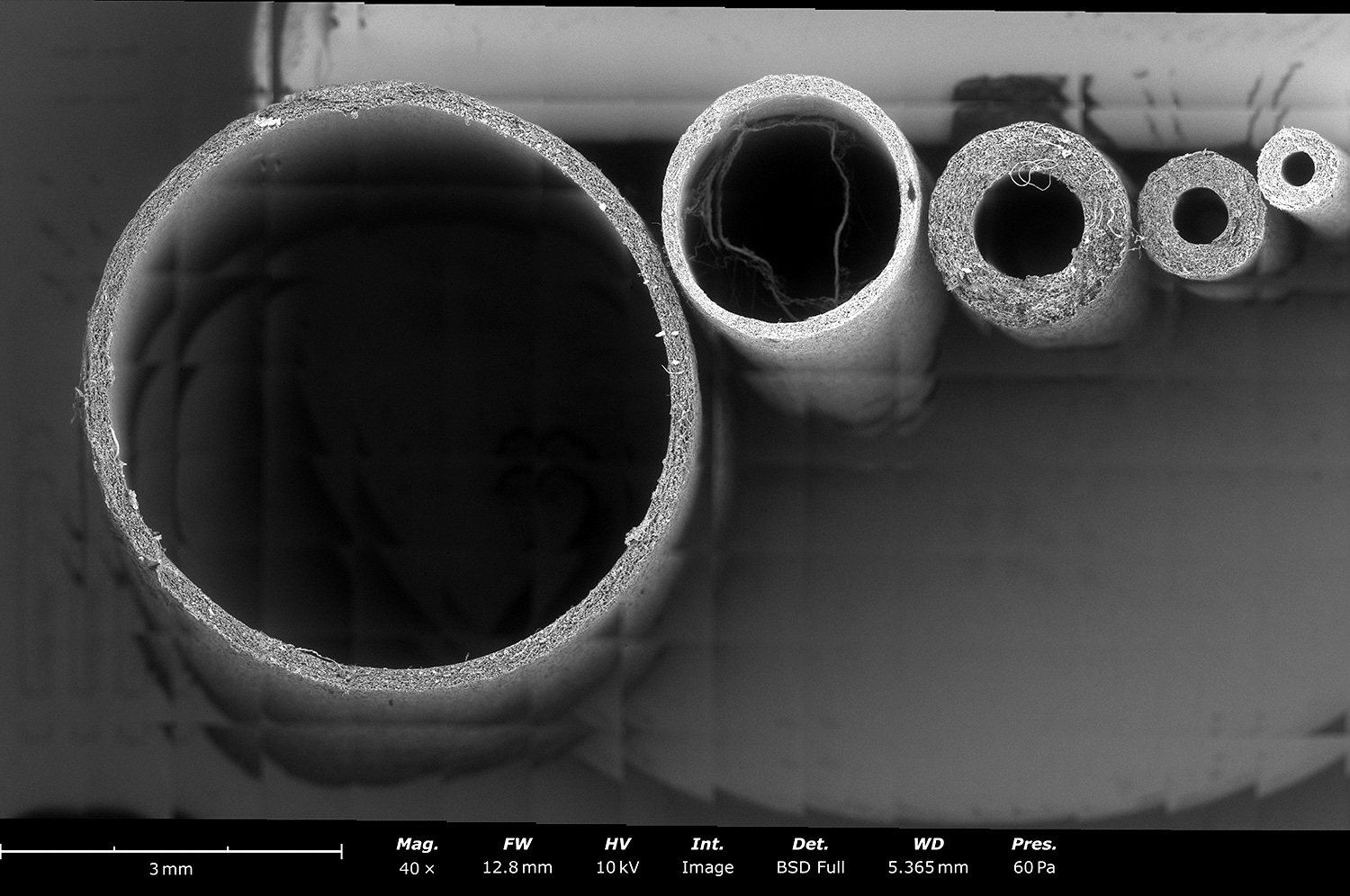
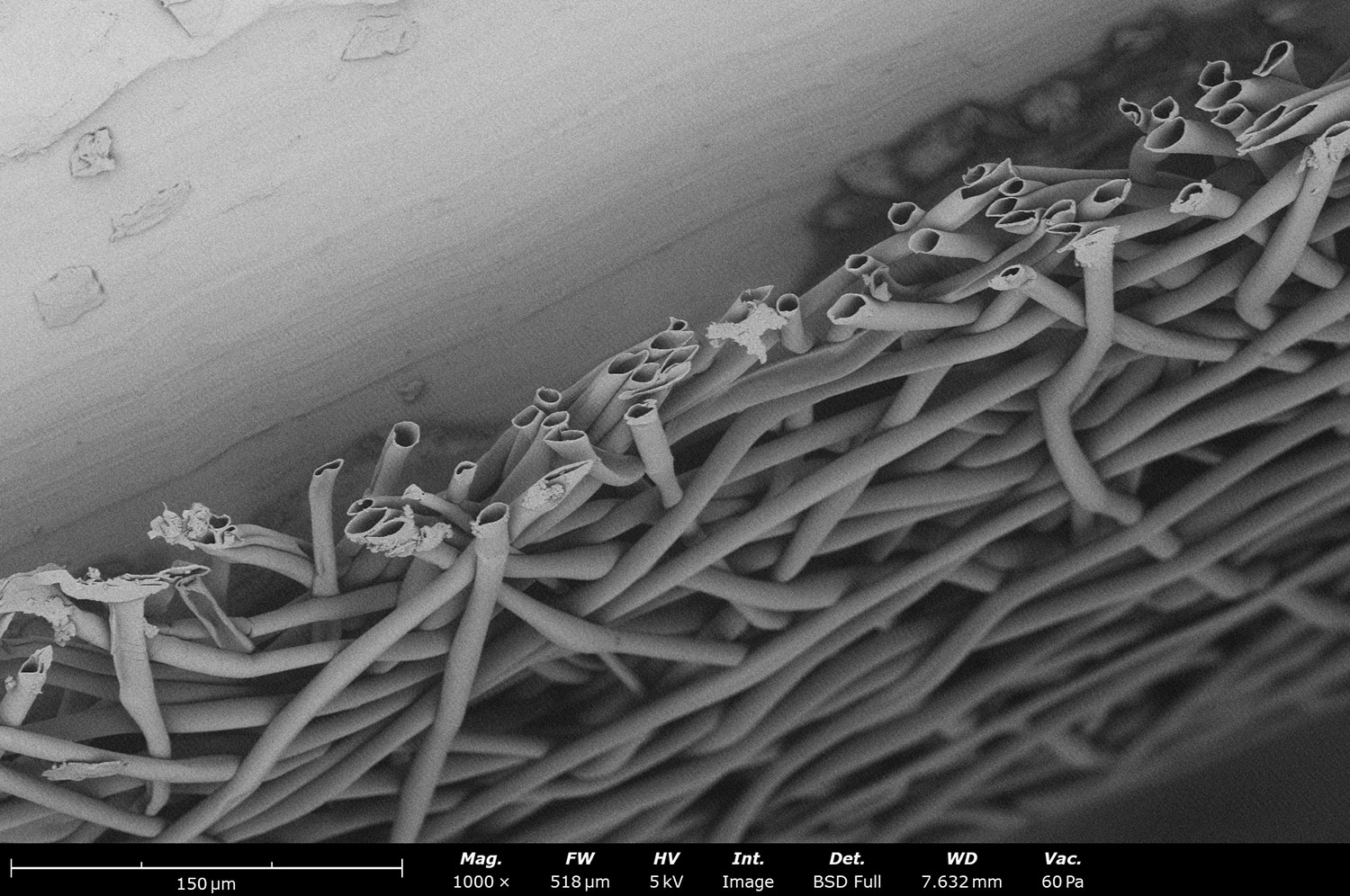
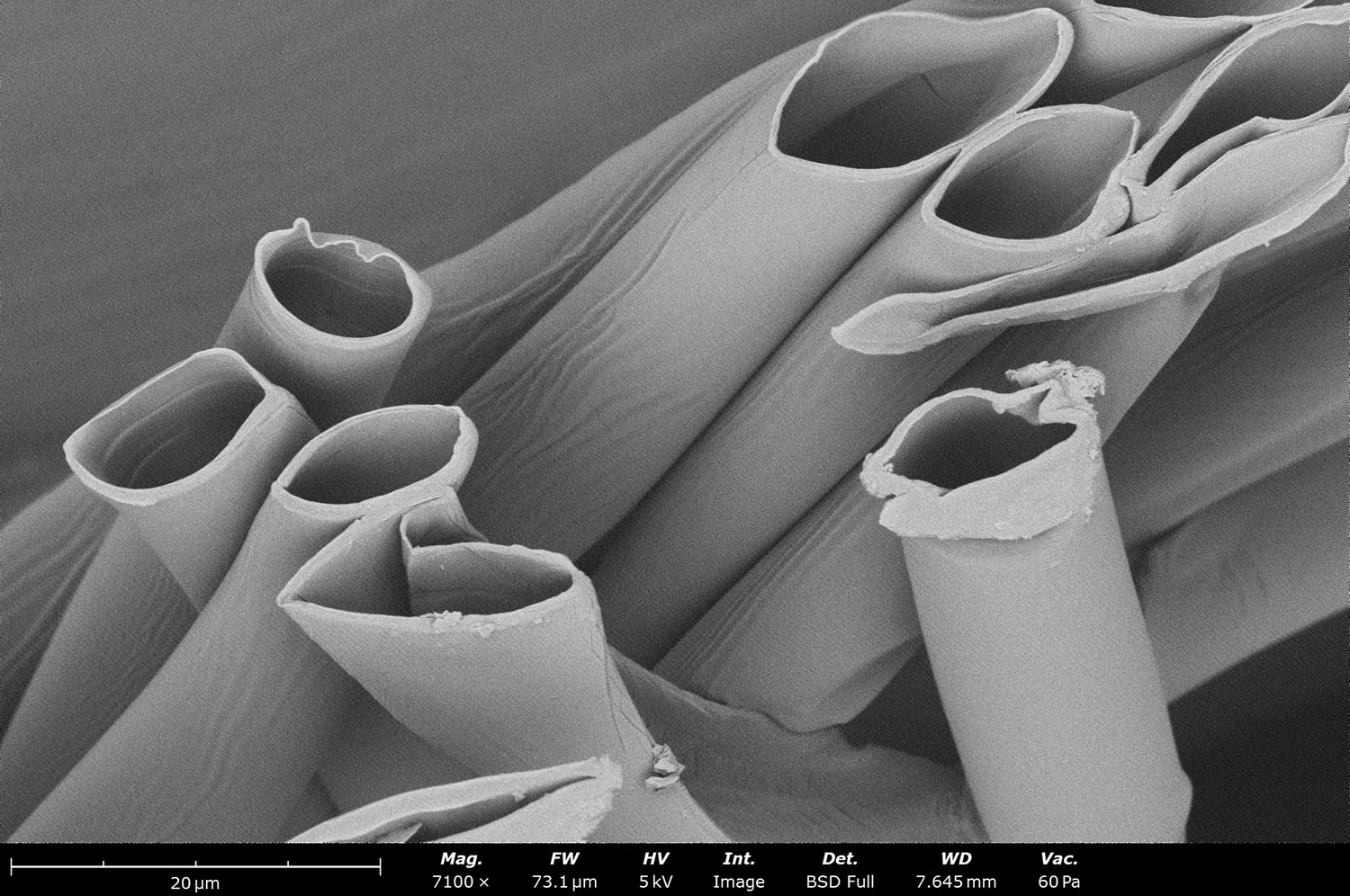
Stitched SEM micrograph showing different electrospun tubular polycaprolactone scaffolds made with the Fluidnatek LE-100 BioTubing. The BioTubing equipment allows to generate tubular structures from less than 1 mm in diameter and up to 20 cm in diameter. These types of samples are commonly used as artificial blood vessels with different polymers, fiber orientations, diameters and even multi-layered structures.
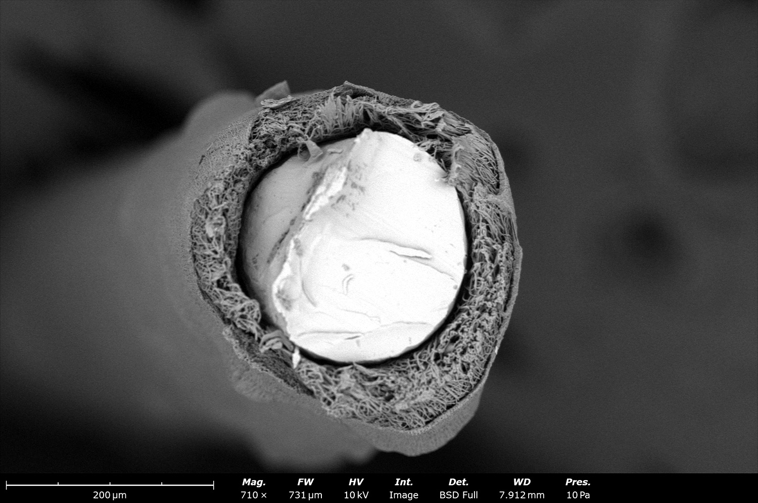
Backscattered electron micrograph of a tubular structure made out of polycaprolactone (PCL) with the Fluidnatek LE-100 BioTubing. The cross-sectional image was obtained by freeze fracturing PCL with liquid nitrogen. If cut at room temperature, the sample will smear, and the microstructure was not going to remain intact as its glass transition temperature is -60°C.
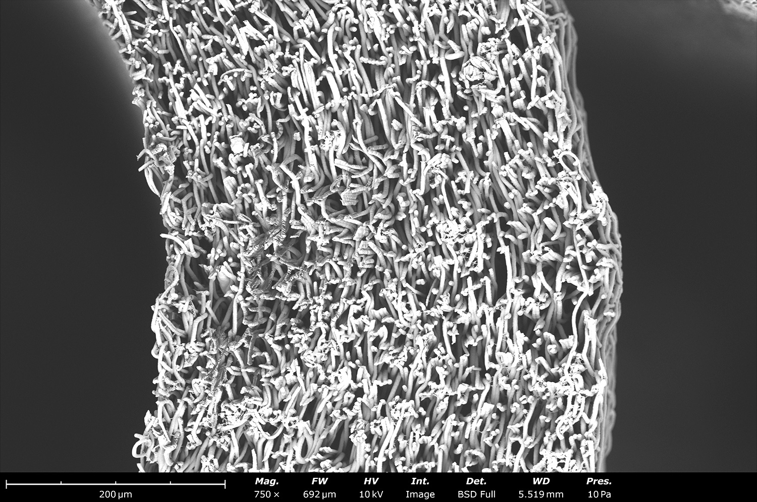
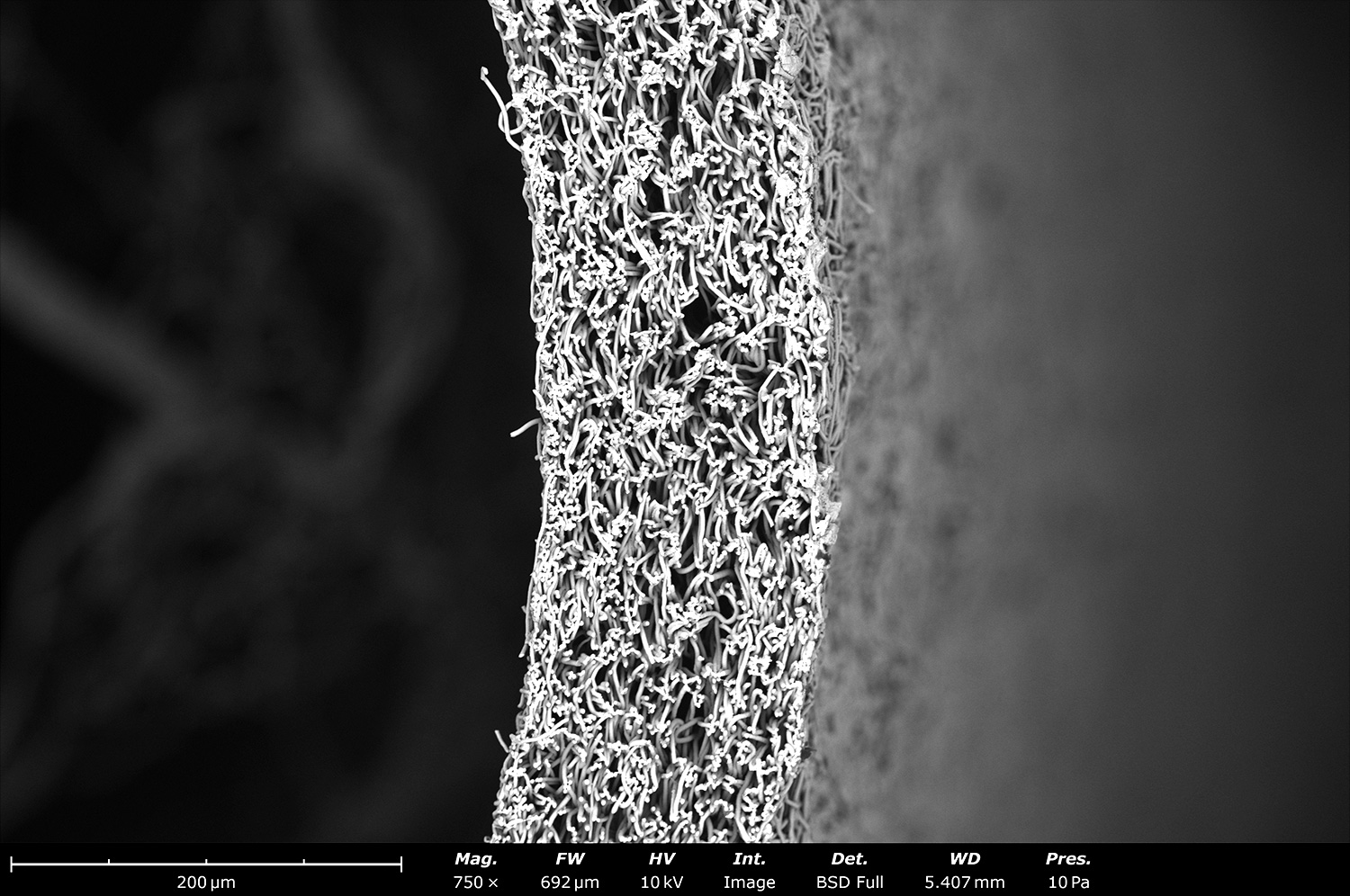
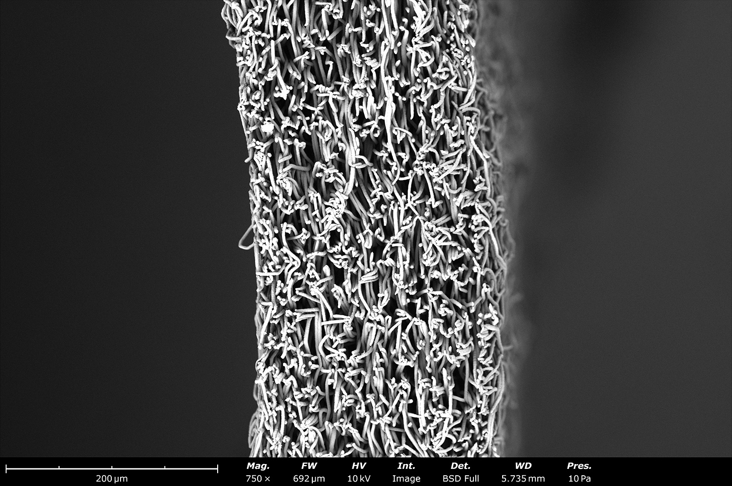
This image shows the cross-section of electrospun polycaprolactone fibers collected on a 2 mm diameter rod with the Fluidnatek LE-100 BioTubing. Sample thickness was approximately 150 µm and homogeneous across its diameter and length. Artificial blood vessels up to 40 cm in length can be obtained with the Fluidnatek technology, or longer upon request.
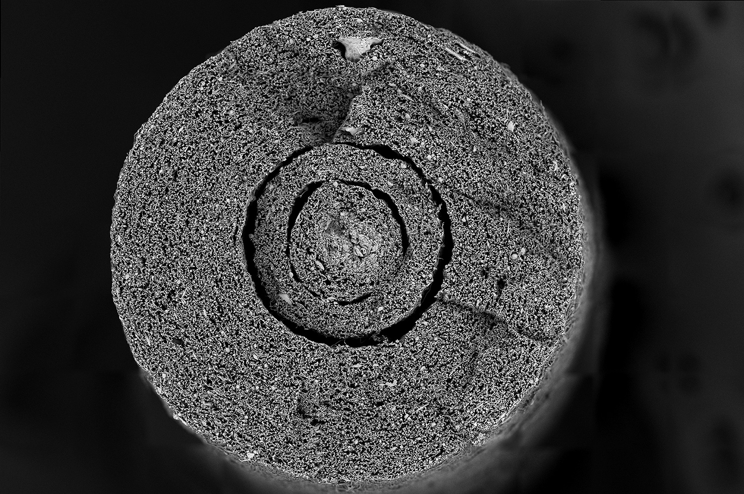
This SEM micrograph shows the cross-sectional structure of polycaprolactone fibers collected onto a 5 mm diameter rod with the Fluidnatek LE-100 BioTubing. The sample is completely porous and if needed, two materials can be collected at the same time to improve mechanical properties, tune porosity and pore size, or have additives like vascular growth factors.
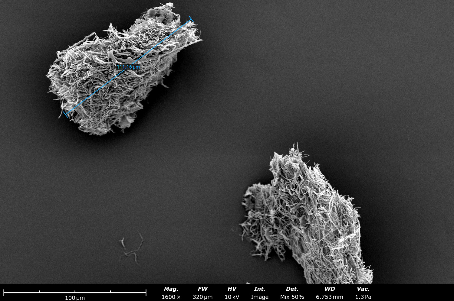
Electron micrograph of micronized scaffold electrospun nanofibers made from a natural protein. Electrospun fibers can be engineered to biomimic the human extracellular matrix. By micronizing the electrospun scaffold these micronized nanofibers can be applied to complex or irregular topography for wound healing. This image was acquired by mixing the signal of the backscatter and secondary electron detectors at a 1:1 ratio.
This SEM image shows a closer look of micronized electrospun nanofibers made from a natural protein. While the micronized sample has a length less than 150 µm, the fiber diameter is less than 450 nm in diameter. These micronized fibers can be fully engineered and can be applied dry or hydrated based on the targeted tissue.
Coronary artery stent coated with electrospun thermoplastic polyurethane (TPU). Electrospinning allows direct deposition onto a metal, or non-metal, mesh to improve biocompatibility of the medical devices. Adhesion and bonding of electrospun fibers onto the mesh can be achieved and will withstand tears during crimping. This process reduces manufacturing cost as it does not need suturing by hand.
Backscattered electron micrograph of ribbon shaped electrospun fibers made out of gelatin type A from porcine skin. Gelatin fibers are commonly used in the medical field and can be found on products approved, and cleared, by the Food and Drug Administration (FDA). Gelatin from other sources, like fish and bovine, are also processed to generate electrospun fibers or electrosprayed particles.
This SEM image shows the microstructure of electrospun silk fibroin (SF), which is typically used in applications like tissue engineering and drug delivery. Silk fibroin is typically electrospun by using a secondary polymer to makes SF electrospinnable. In this case, polyethylene oxide (PEO) was used as the additive polymer to generate the scaffolding material.
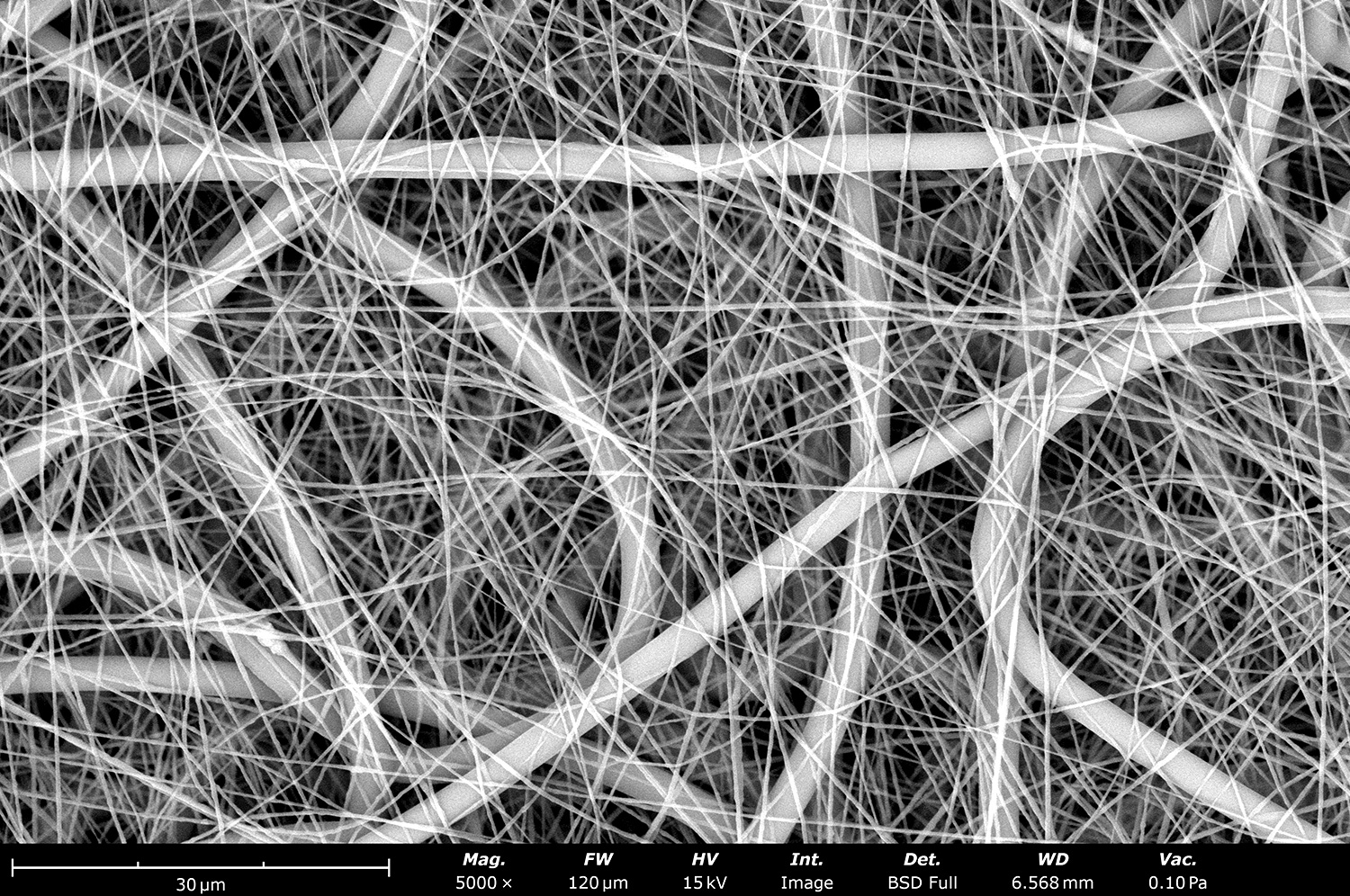
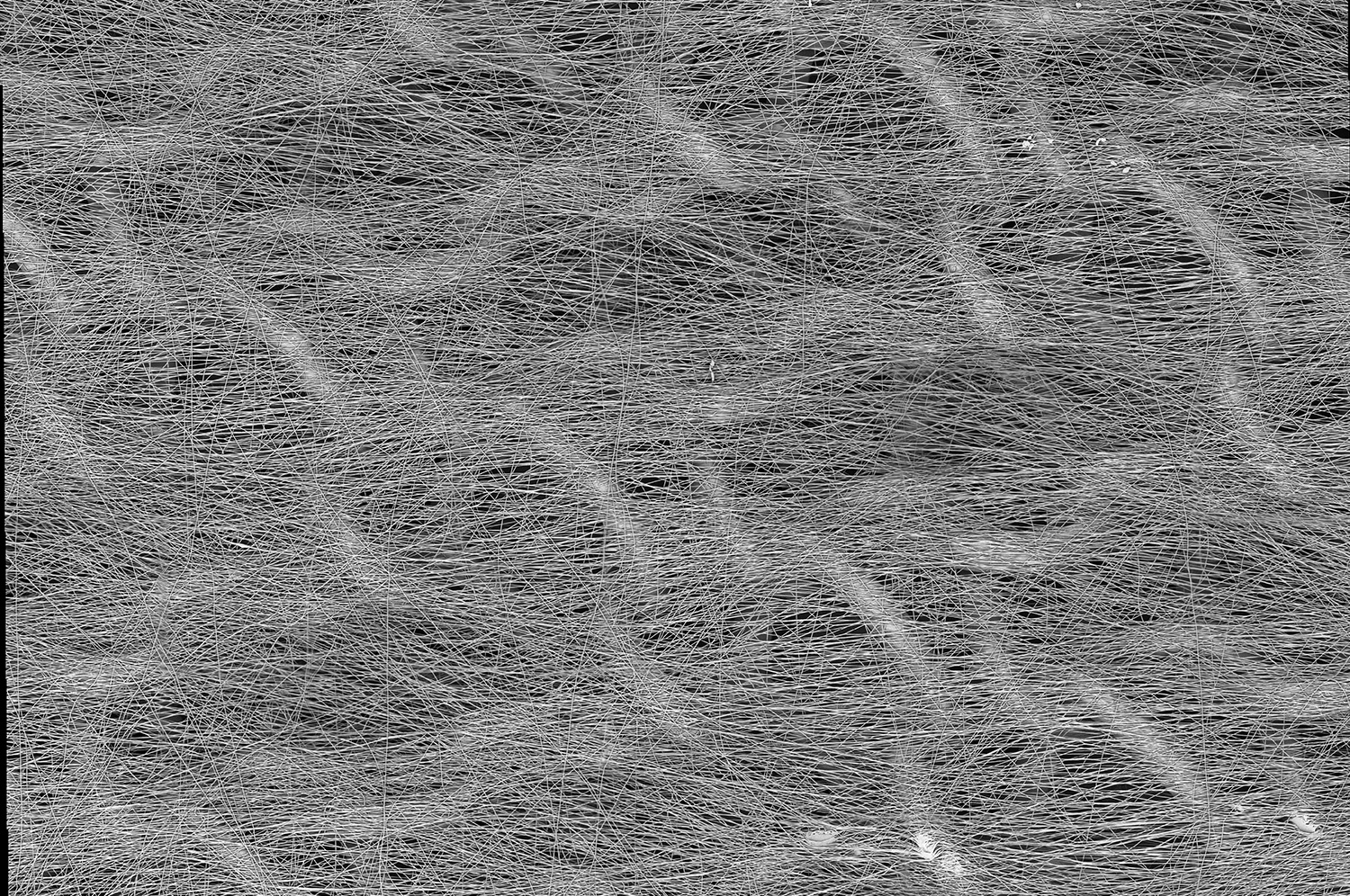
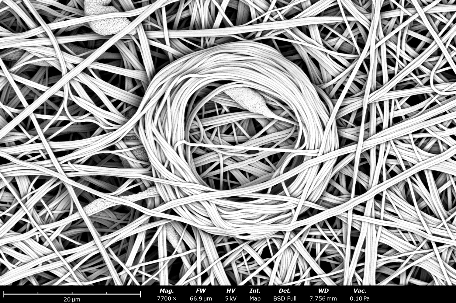
Microstructure of electrospun silk fibroin (SF) with nanofibers from webbing. Webbing is a common defect on electrospun fibers that can occur when the processing parameters have not been optimized. This defect is commonly seen when operating at low relative humidities where the polymer solution can evaporate quickly, causing the formation of these nanoscale fibers with diameter below 100 nm.
This SEM image shows the microstructure of poly(vinylidene fluoride-co-hexafluoropropylene) (PVDF-HFP) containing inorganic particles. Electrospinning offers the capability to generate fibrous materials containing different types of additives to improve sample properties based on application needs.
This microstructure image shows polycaprolactone (PCL) electrospun fibers with a bumpy structure. This rougher microstructure can be engineered for filtration applications where improvement of filtration capabilities is needed for filters made from nanoscale fibers. This image was obtained from a field of depth over 600 µm on a tilted sample with the aid of a python script using the Phenom Desktop SEM.
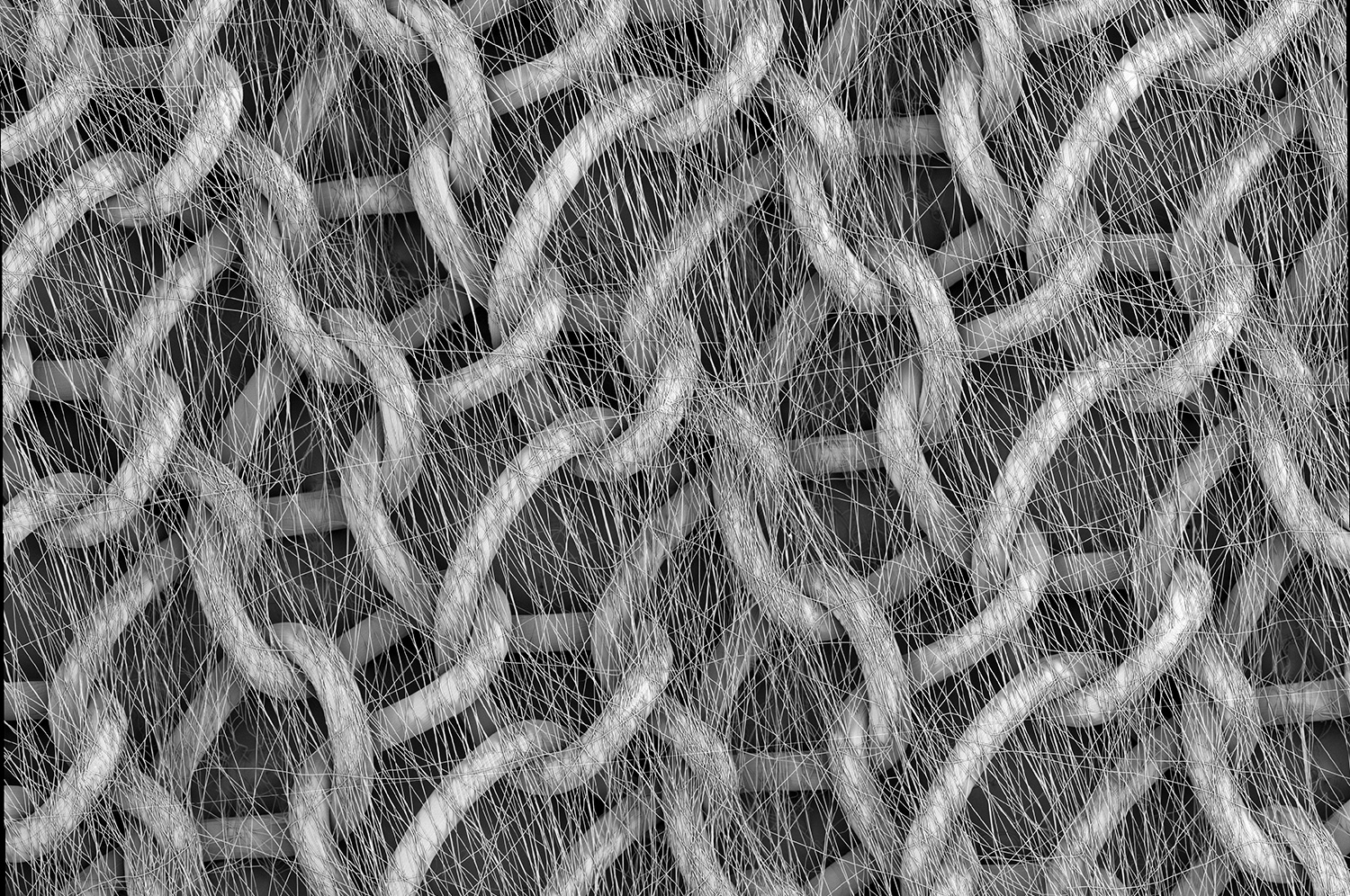
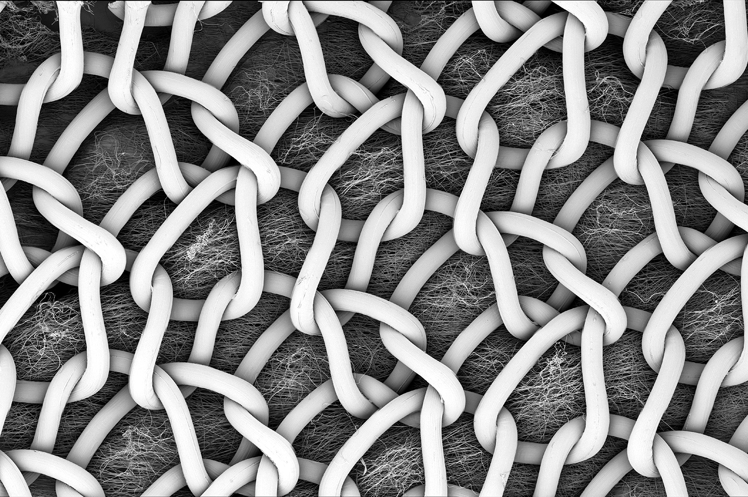
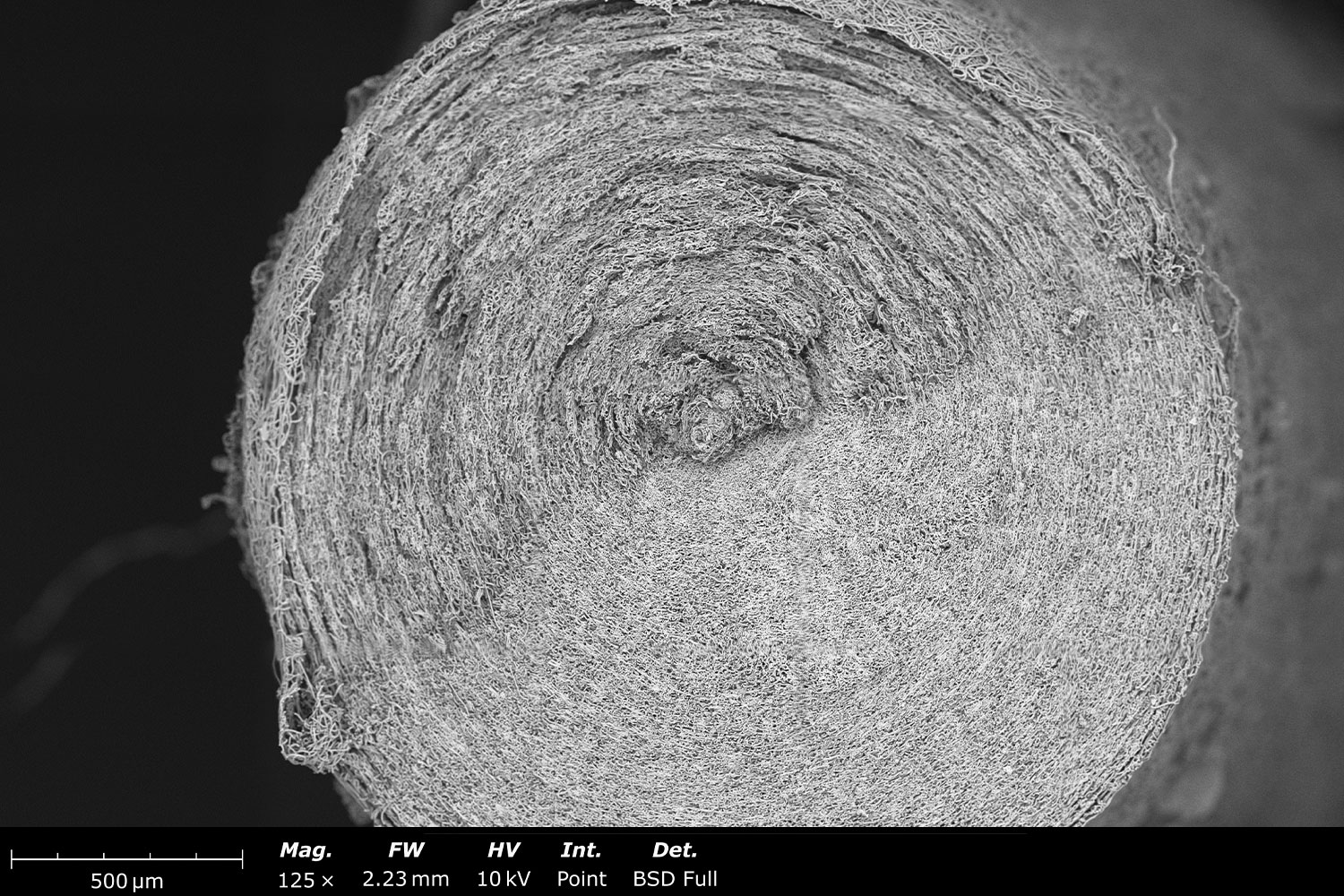
This stitched image shows the microstructure of two electrospun polyhydroxybutyrate (PHB) based yarns made with the Fluidnatek technology. Yarns made out of electrospinning offer the innate ability to resemble the extracellular matrix, incorporate additives like drugs, generate complex structures from two different types of fibers, or even combine fibers and particles on the same yarn.
This close up microstructure of the electrospun PHB yarn shows the degree of twist on the final sample. Yarns developed with electrospinning can be used for suture based applications, whether from pure electrospun fibers, or by reinforcing a yarn with nano- or micro-scale fibers to improve mechanical properties needed during the suturing process. Once the yarn process is optimized, more than 30 meters can be developed in one run with our yarn collector.
Microstructure of electrospun polyacrylonitrile (PAN) imaged using a backscatter electron detector with the Phenom Pharos desktop SEM. PAN is commonly used for hot gas filtration applications as it offers good thermal stability and mechanical strength needed. This sample was stabilized at a temperature of 250°C before imaging. Oxidative stabilization is needed prior to the carbonization step.
This SEM image shows carbonized polyacrylonitrile (CPAN) obtained after heat treatment at temperatures above 1,000°C under inert environment. CPAN has excellent electrical conductivity and offers good mechanical strength needed when used for fuel cell applications. Note the reduction in fiber diameter from its stabilized predecessor while still maintaining a homogeneous diameter post-carbonization.
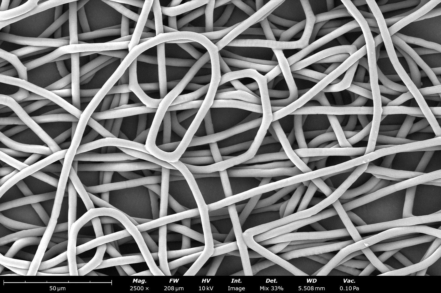
This backscattered electron micrograph shows the effect on the microstructure of Polycaprolactone (PCL) electrospun fibers when a low voltage (-5 kV) is used on the collector in a Fluidnatek LE-100. Since the fibers have more attraction towards the collector (when compared to a grounded collector) they have less time to stretch during the process, creating the curly pattern throughout the sample. Fiber-fiber bonding between fibers is also increased as the solvent does not have enough time to fully evaporate.
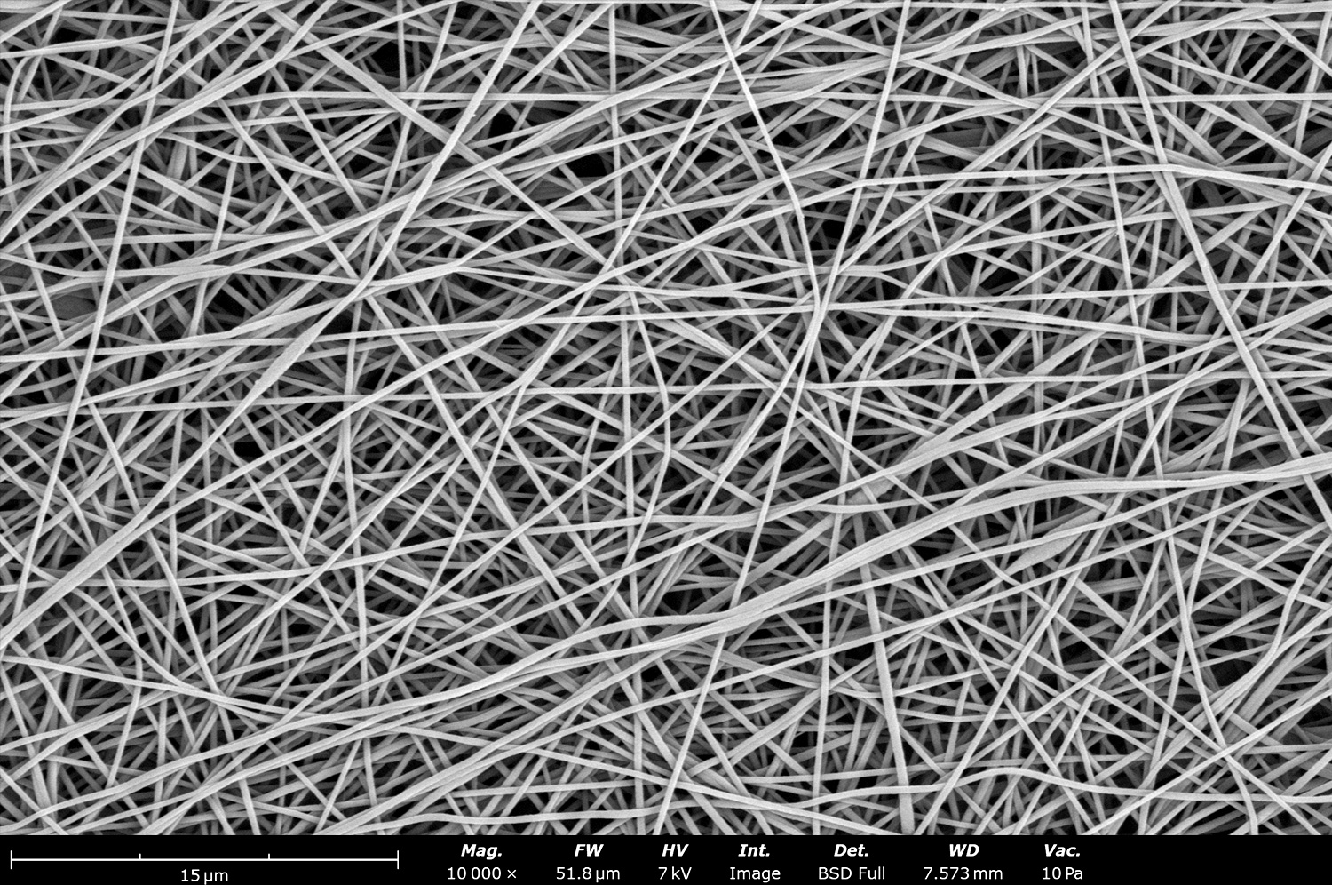
Micrograph image of thermoplastic polyurethane (TPU) microfibers generated with the Fluidnatek LE-50 using a drum collector of 10 cm in diameter. Electrospun TPU fibers are commonly generated with a diameter below 2 µm. By properly optimizing the solvent system the fiber diameter can be adjusted to increase the diameter while maintaining a homogeneous structure as shown on this image.
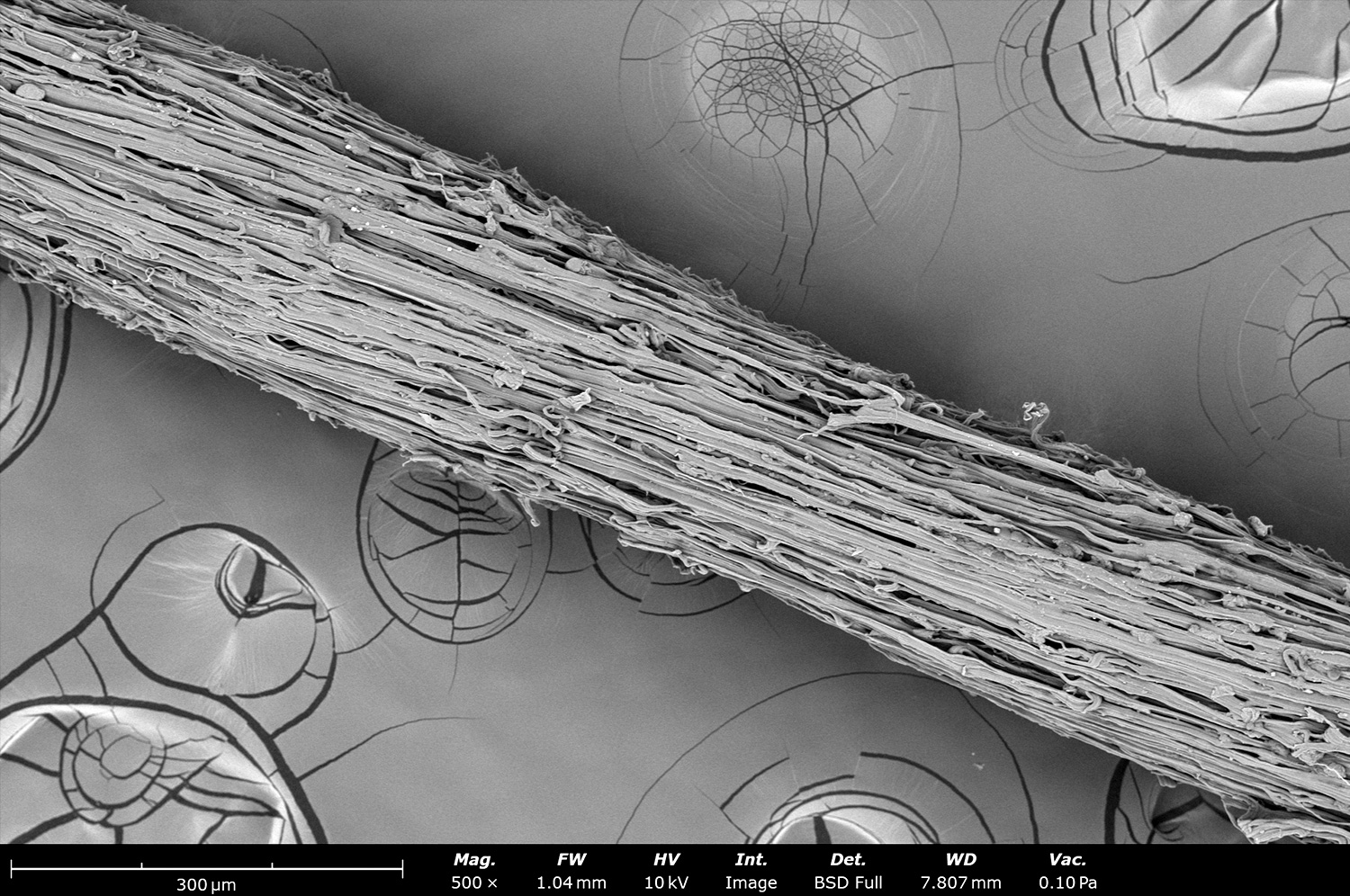
This image shows a close up of the microstructure of thermoplastic polyurethane (TPU) fibers. These TPU fibers have a fiber diameter around 10 µm, allowing the final sample to become significantly flexible while obtaining a large pore size used for cell infiltration. These large fibers are ideal to coat stent, ballons, meshes and other medical devices for implantation.
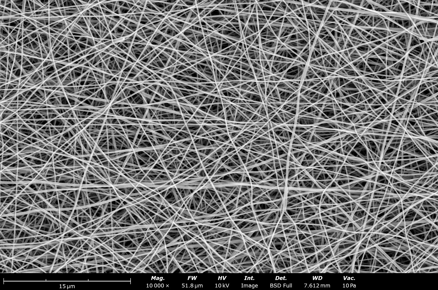
This SEM image shows the microstructure of a thermoplastic polyurethane (TPU) microfibers that was co-electrospun with polyethylene oxide (PEO) nanofibers. The Fluidnatek equipment allows its users to process two different materials with independent voltages. This sample was generated with the Fluidnatek LE-50 under tight temperature and relative humidity.
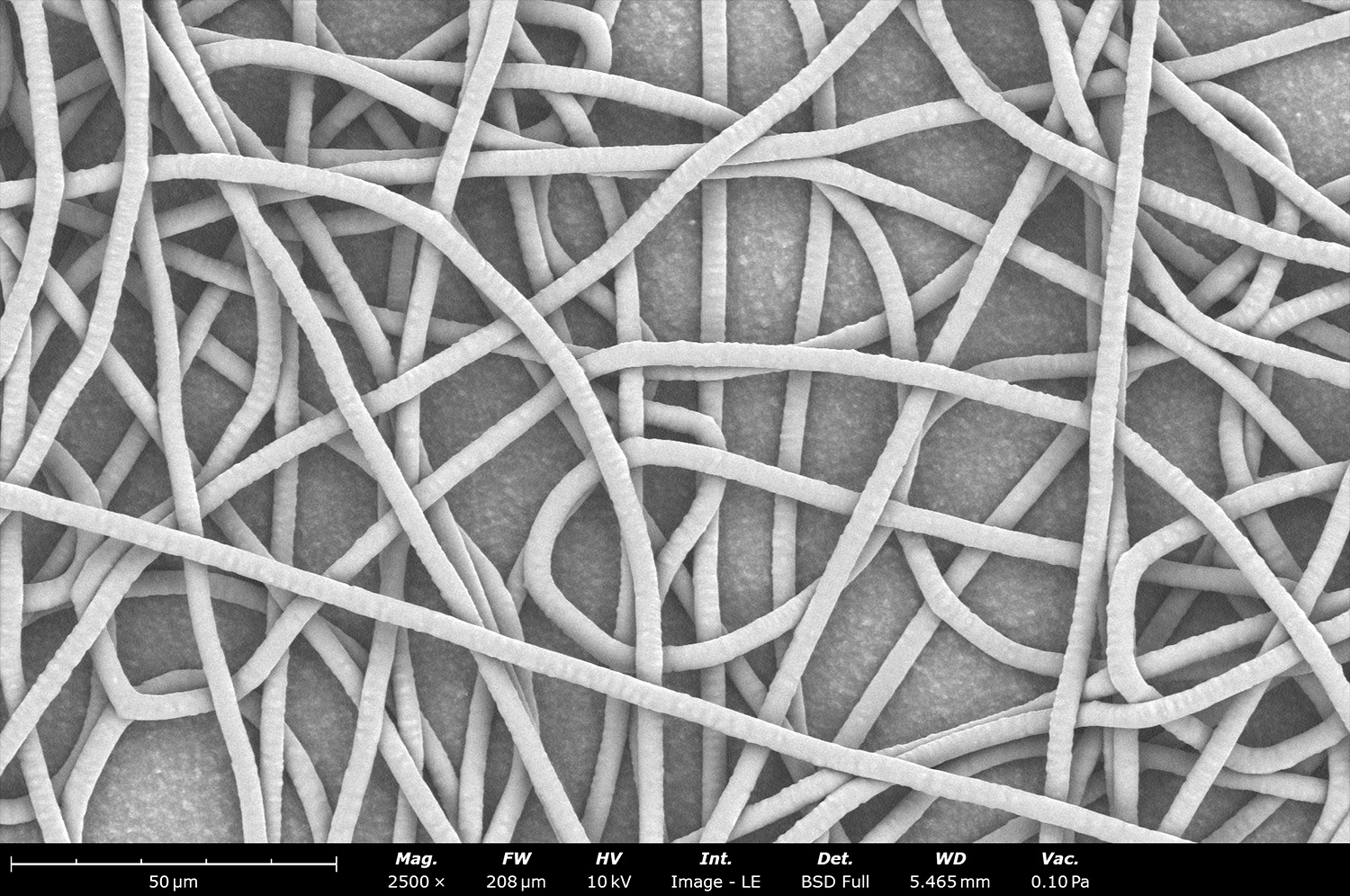
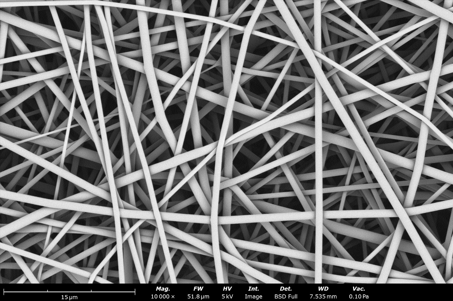
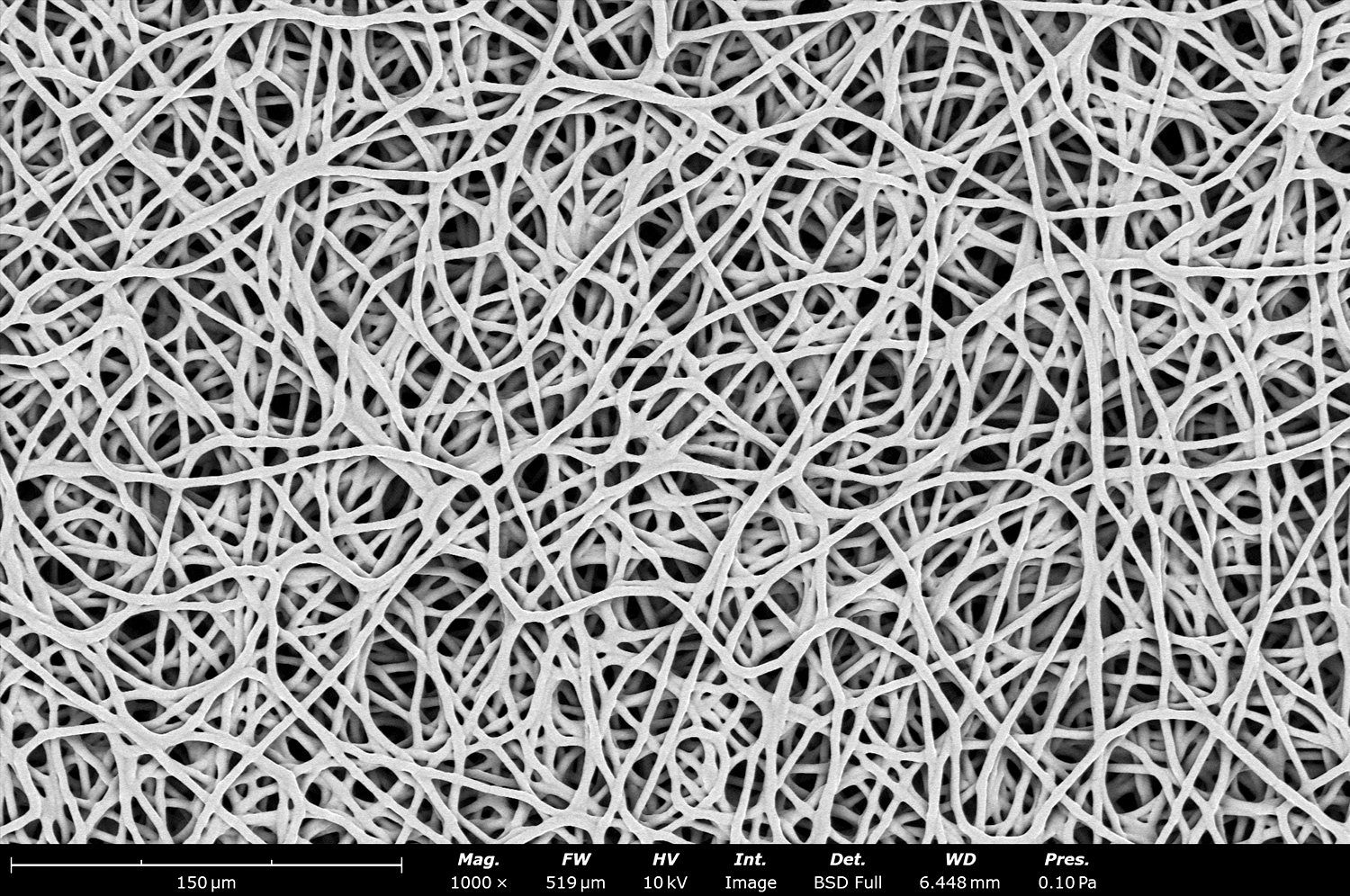
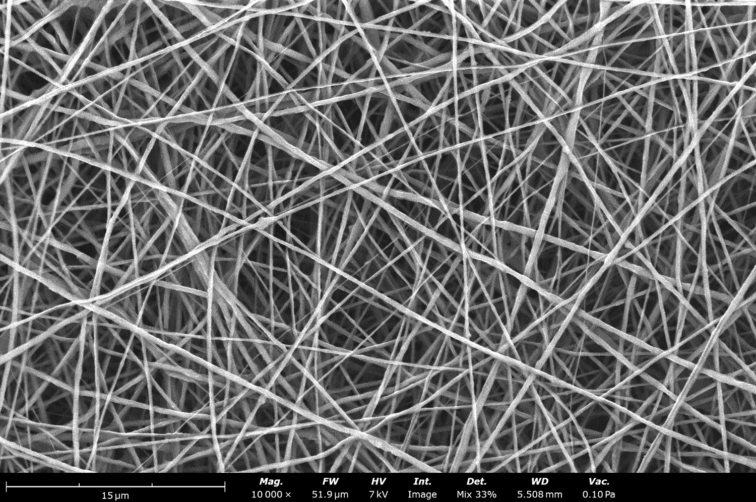
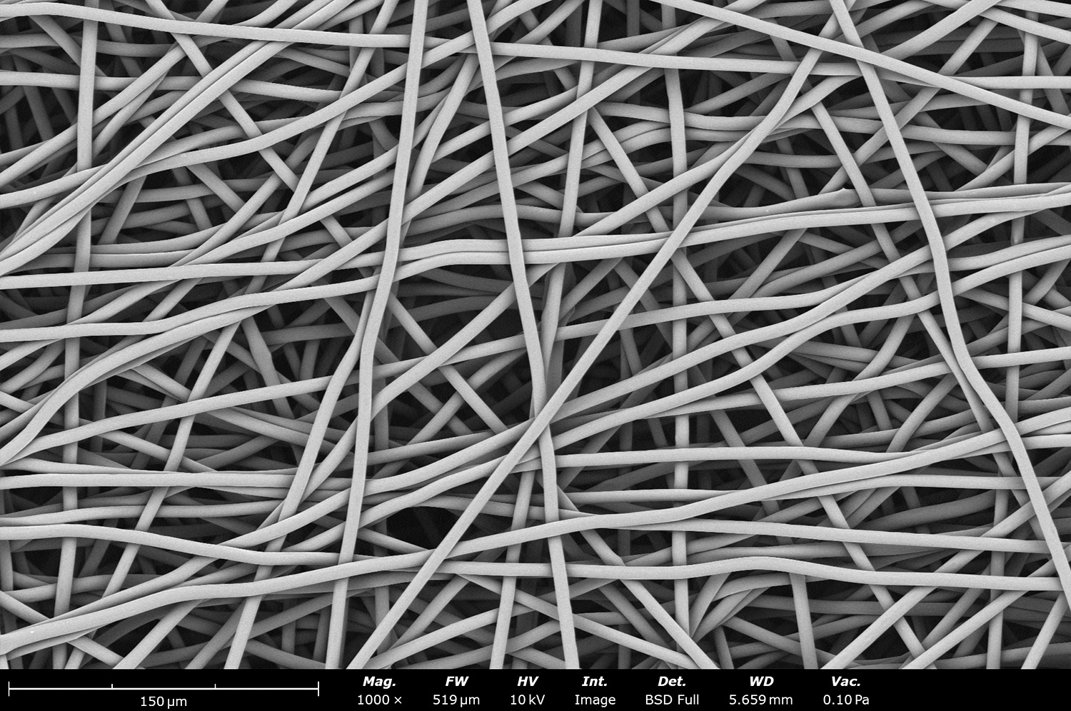
This SEM image shows the microstructure of Polycaprolactone (PCL) fibers made using dichloromethane (DCM) as the solvent under tight temperature and relative humidity with the Fluidnatek LE-50. The image shows a defect free microstructure, showcasing that perfect fibers can be obtained over a large sample area while maintaining a tight fiber diameter.
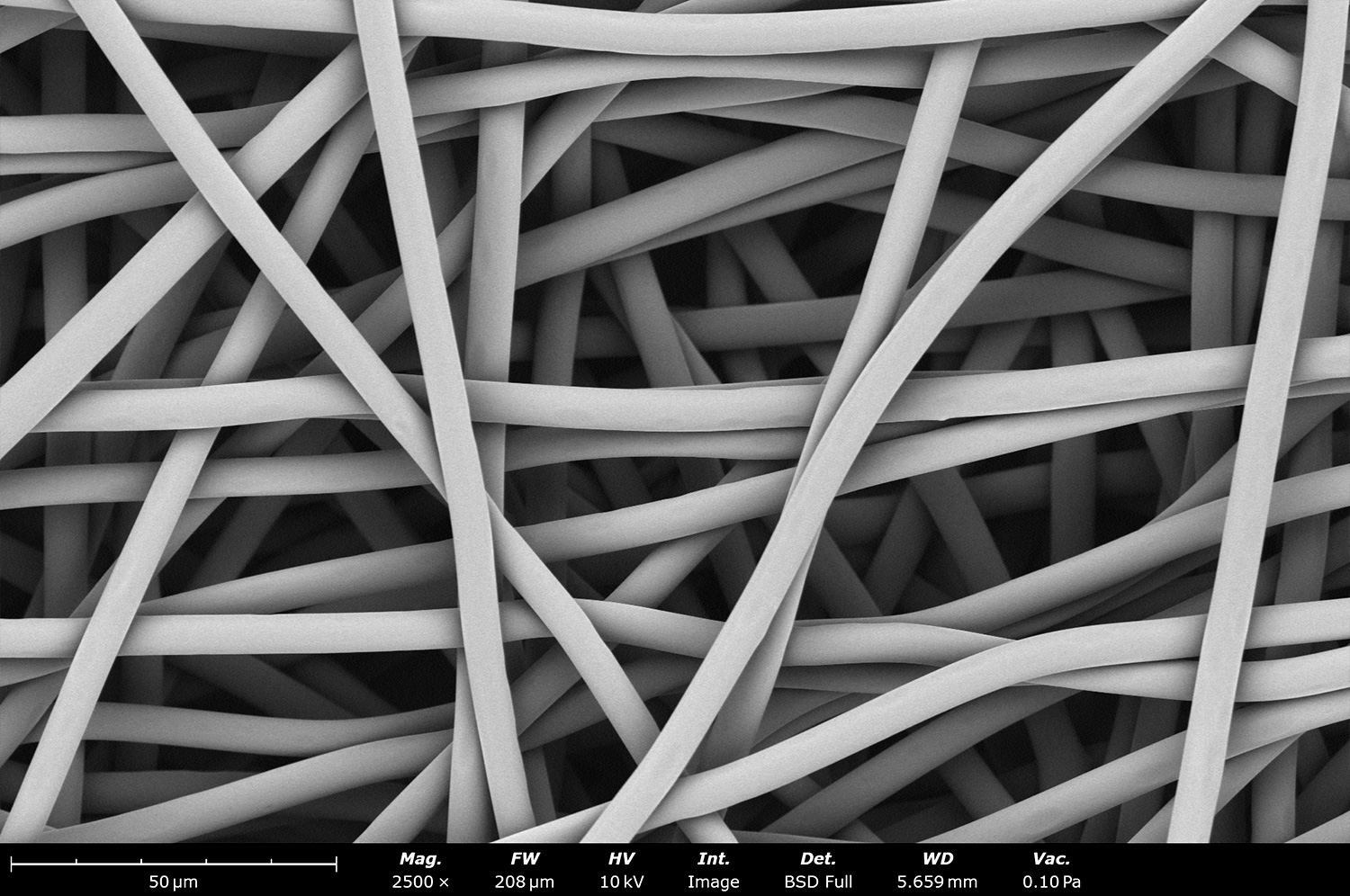
Electron micrograph of porous polycaprolactone (PCL) microfibers with a diameter averaging 10 µm. The uniformity of the fiber diameter and porous structure on the surface is clearly seen. This surface nanoscale porosity significantly increases the surface area of the samples. The nanoscale pores improve cell adhesion and proliferation when used for tissue engineering applications.
Poly(vinylidene fluoride-trifluoroethylene) (PVDF-TrFE) is a synthetic polymer with piezoelectric properties. A piezoelectric material can convert mechanical energy into electrical energy, or the other way around. These electrospun fibers were generated with the Fluidnatek LE-50 and have an average fiber diameter around 780 nm. Solution and processing optimization can be performed to decrease its diameter based on application needs.
SEM image of electrospun poly(vinylidene fluoride-trifluoroethylene) (PVDF-TrFE) nanofibers. In this case, solution optimization was performed to effectively reduce the fiber diameter to 350 nm. Having PVDF-TrFE with an aligned structure can help neurons proliferate easier when compared to randomly oriented fibers.
Poly(vinylidene fluoride-trifluoroethylene) (PVDF-TrFE) electrospun nanofibers with an average fiber diameter below 150 nm. These fibers offer a promising alternative to the alpha and beta crystalline structures typically found with larger fibers. Higher quantities of beta structure can improve piezoelectric properties. Upon close inspection, fibers with an average fiber diameter of 20 nm are seen.
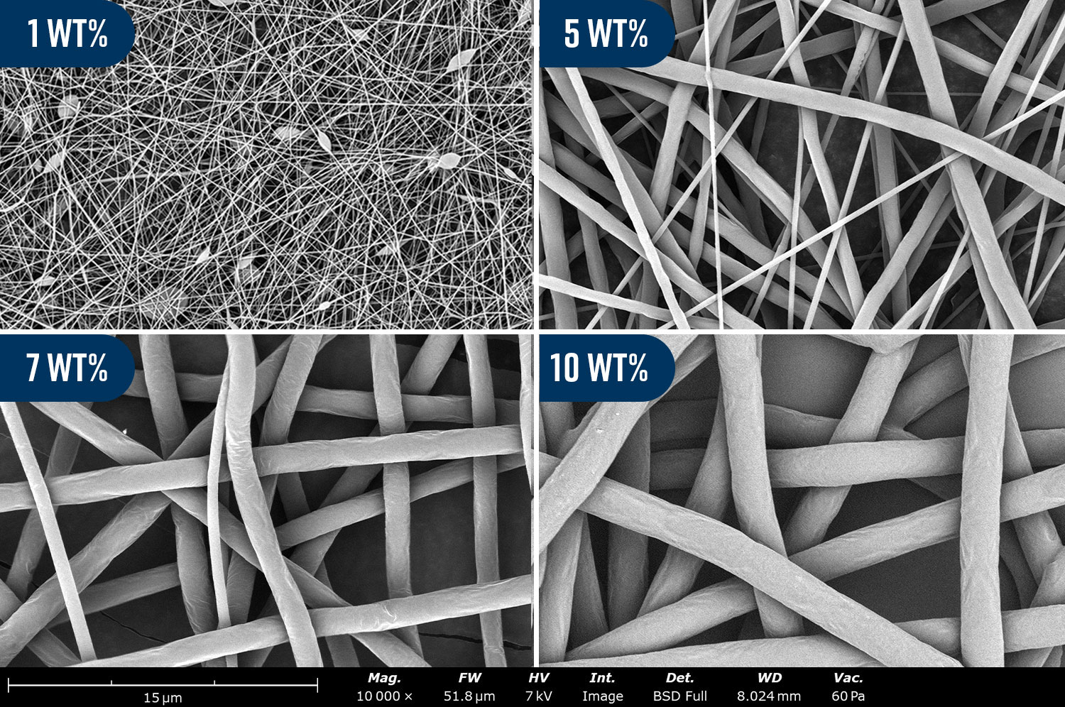
These micrographs showcase the resulting microstructure from different wt% polycaprolactone in hexafluoroisopropanol solution. The 1 wt% solution results in nanoscale fibers with a diameter of 98 ± 29 nm mixed with beaded fibers. Beads are typically not desired in electrospinning processes and are due to low concentrations and low viscosities. The 5 wt% solution resulted in the formation of fibers without beads with a fiber diameter of 942 ± 522 nm. A further increase to 7 wt% and 10 wt% solutions resulted in increases in fiber diameter to 1.81 ± 0.63 µm and 2.35 ± 0.10 µm respectively. Larger fiber diameter is also associated with an increase in pore size. These larger and more uniform fibers can be used in applications like tissue engineering and coating medical devices.
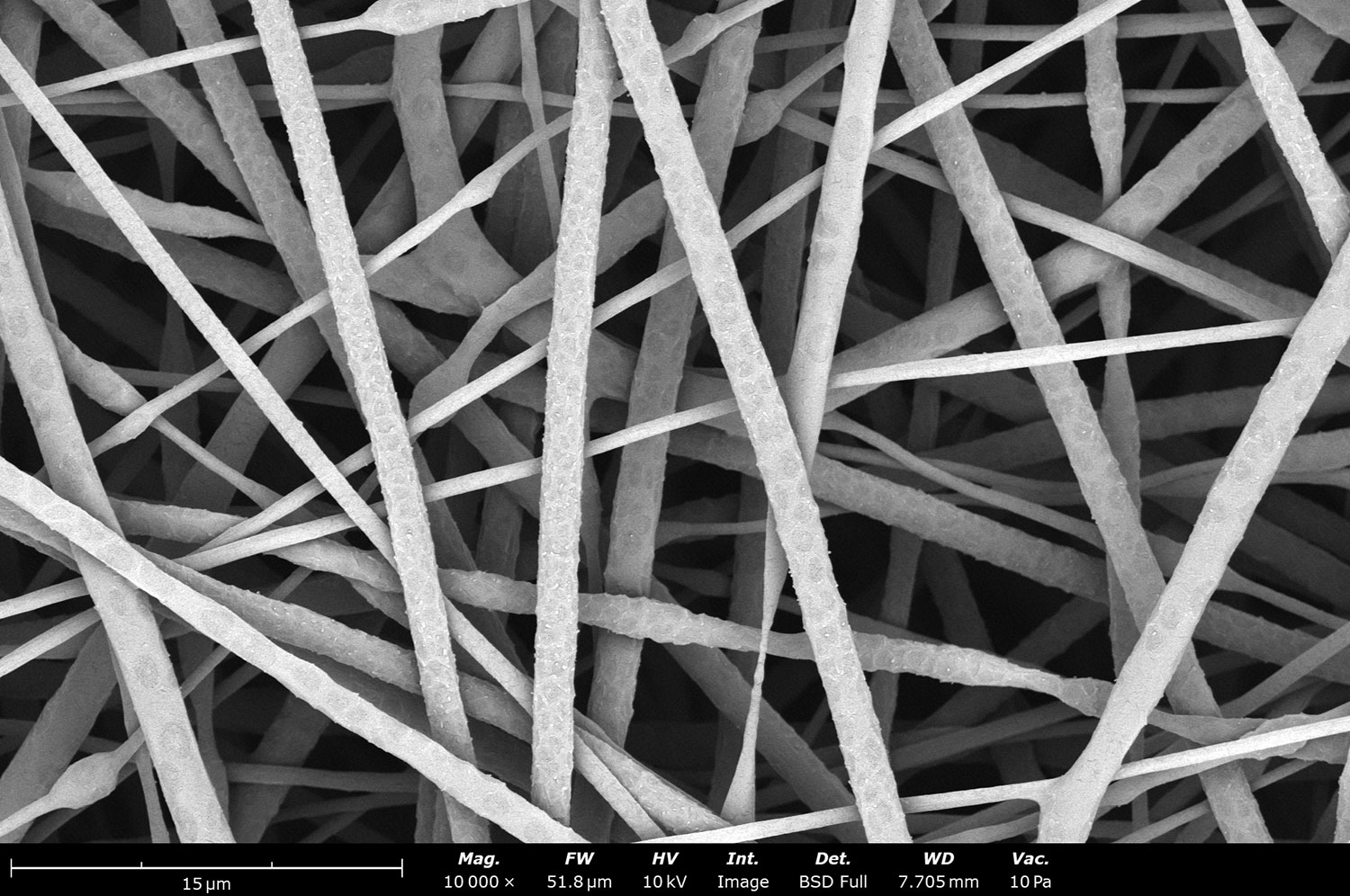
This microstructure showcases a 10 wt% polycaprolactone in hexafluoroisopropanol solution containing an extra 10 wt% of organic salt based on polymer content. The incorporation of the organic salt has increased the solution conductivity, allowing the fiber to reduce from 2.35 ± 0.10 µm to 1.02 ± 0.50 µm. Residual salt is also observed in the surface of the fibers.
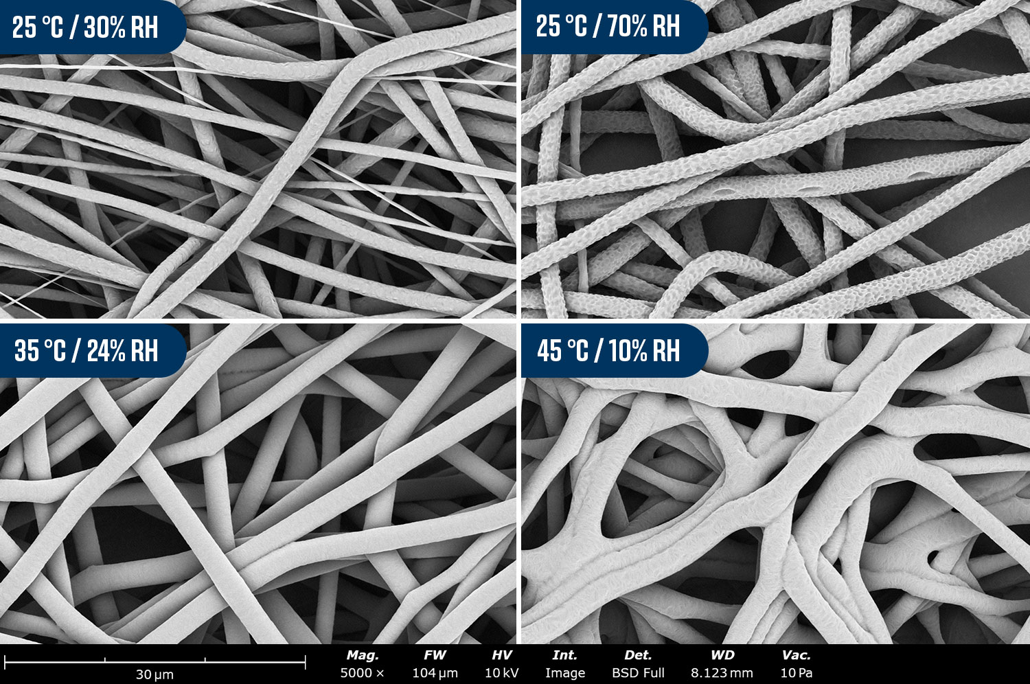
These images show the microstructure of electrospun fibers processed with a 13.5 wt% polycaprolactone in chloroform:methanol (10:1) solution at different temperatures and relative humidities. While at 25°C/30% the fibers have bimodal diameter, at 25°C/70% high surface porosity is seen. Optimizing to 35°C/24% allows uniform fiber diameter. Adjusting to 45°C/10% has a negative effect on fiber microstructure and generates significant quantities of fiber-fiber bonding.
This electron micrograph shows a closer look at the porous structure on the polycaprolactone electrospun fibers. These large fibers become flexible, a key aspect for various applications needing a resilient sample that can withstand mechanical compliance for samples that need to expand and retract. A common sample where these fibers are used are artificial blood vessels for vascular applications.
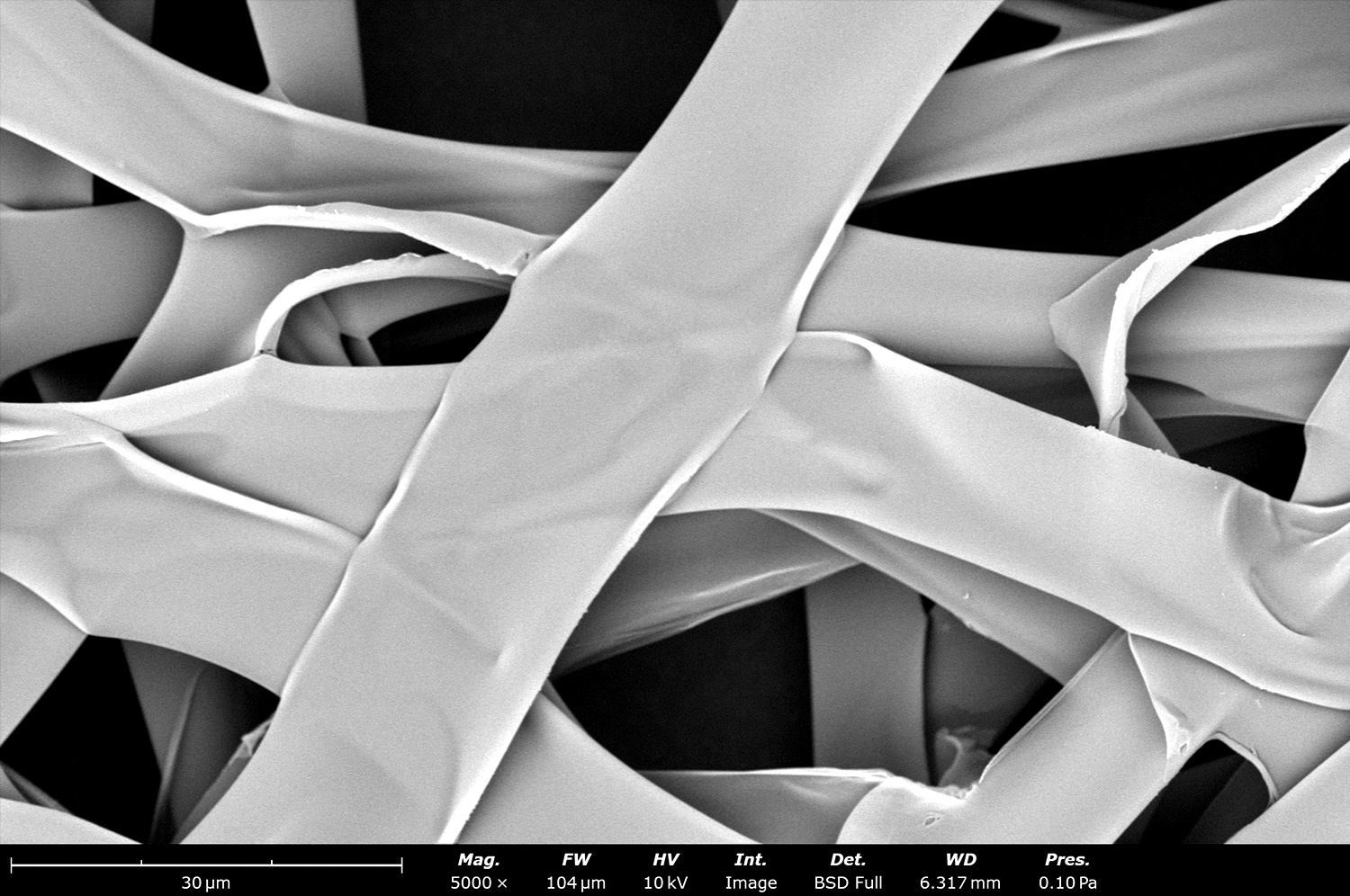
This image showcases the microstructure of hollow electrospun fibers made with polylactic acid (PLA). The resultant morphology was able to be obtained by processing the polymer in coaxial mode with PLA in the shell and mineral oil in the core. The sample was then post-processed to leach out the mineral oil, resulting in the hollow structure shown here.
Hollow fibers have a wide range of uses for different application needs. Two common applications are the encapsulation of additives that can be released over time, and to capture carbon dioxide from the environment. The image shown showcases hallow electrospun fibers made with polylactic acid.
Densified cylindrical electrospun scaffolding sample made out of blended polyethylene oxide and polylactic acid fibers. These types of sample morphology is useful in applications like tissue engineering, specifically for tendon repair as the morphology and fiber orientation could help mimic the structure of the native tissue.
This site is protected by reCAPTCHA and the Google Privacy Policy and Terms of Service apply.