Phenom Scanning Electron Microscope
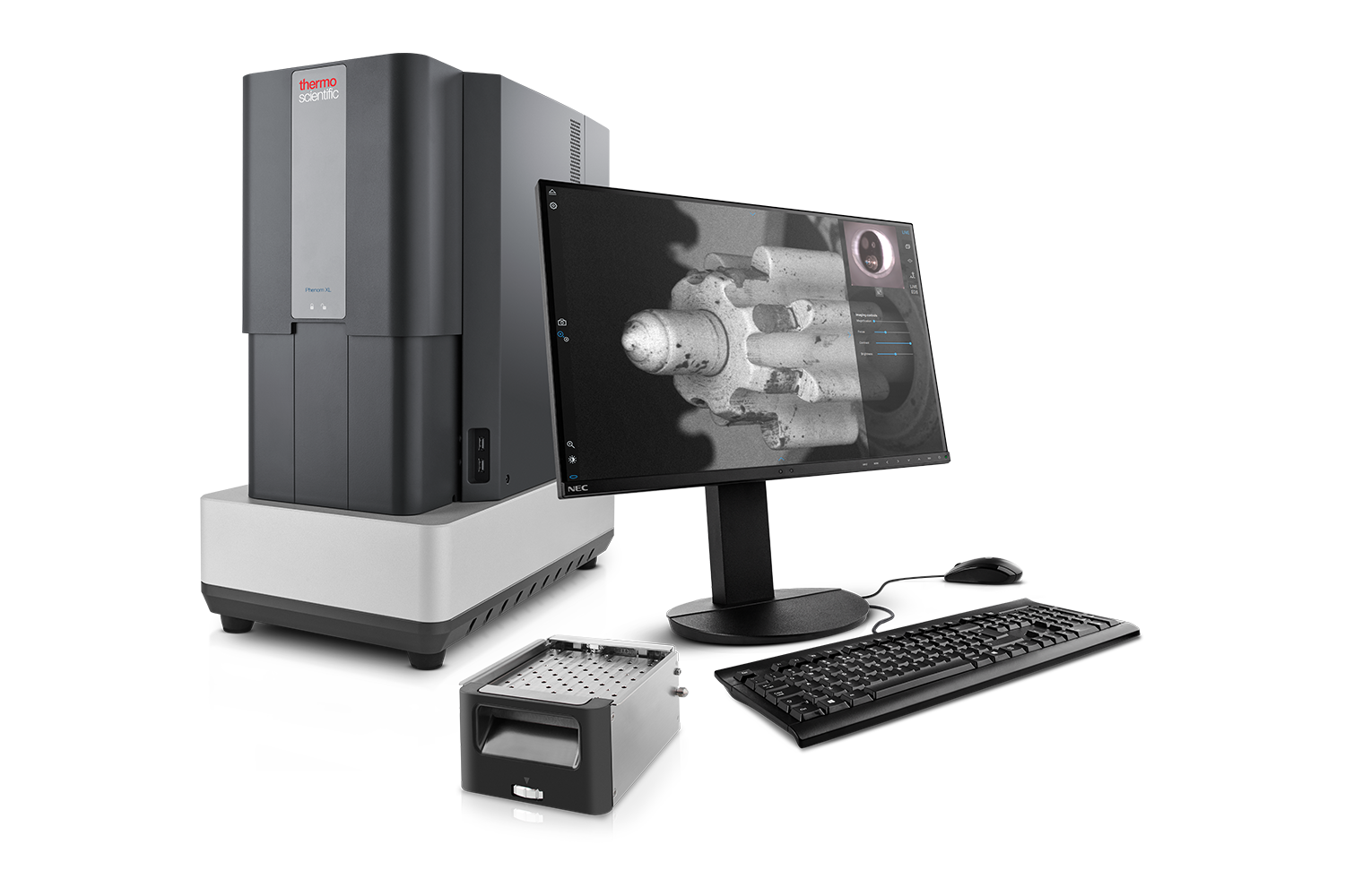
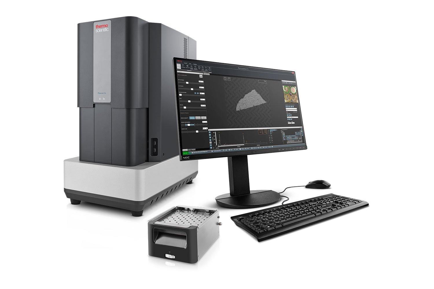
Phenom XL Desktop SEM
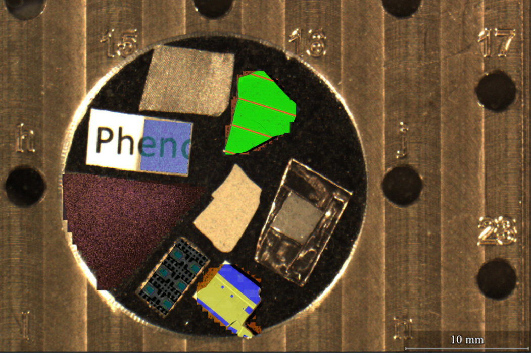
The Phenom XL is a powerful scanning electron microscope (SEM) that combines large sample capacity with an ultra-fast loading mechanism to maximize analysis throughput. It exceeds standard sample size limitations by supporting up to 36 half-inch SEM sample stubs on a 100 mm x 100 mm motorized stage, enabling efficient and versatile sample handling.
Largest Sample Stage
Load up to 36 half-inch SEM stubs in a wide analysis area up to 100 mm x 100 mm.
Automated Imaging & Analysis
Leverage pre-built or custom Python scripts to automate imaging. Integrated Phenom ProSuite for automated image analysis.
Specialized Sample Stages
Motorized eucentric tilting and rotating, tensile testing, vacuum transferring, and resin mounting.
Talk to an Instrumentation Specialist Today!
Best Selling Desktop SEM Empowered with Large Sample Stage
The Phenom XL G3 desktop SEM features the largest sample stage of any desktop SEM and delivers intuitive, automated solutions that reduce manual repetitive tasks. The large chamber enables innovative solutions such as motorized eucentric sample tilting, in situ tensile testing, and electrical feedthrough capabilities. Dry-room and argon compatibile models are available for placing the SEM in inert environments and protecting sensitive samples.
The Phenom XL comes equipped with PyPhenom, a comprehensive Python library that provides users the flexibility to script customizable and automated workflows. It enables the design of personalized GUIs, EDS analysis procedures, and more, enhancing control and efficiency in imaging operations.
Model: |
Phenom Pharos |
Phenom XL |
Phenom Pro/ProX | Phenom Pure |
|---|---|---|---|---|
Specifications |
||||
| Electron Source | Field Emission Gun (FEG) | CeB6 crystal | CeB6 crystal | CeB6 crystal |
| Max. SEM Magnification | 2,000,000x | 200,000x | 350,000x | 175,000x |
| SEM Resolution – BSD | 3 nm | 9 nm | 8 nm | 15 nm |
| SEM Resolution – SED | <2 nm | 9 nm | 6 nm | 15 nm |
| Acceleration Voltages | 1 – 20 kV | 1.5 – 20 kV | 1.5 – 20 kV | 2, 5 or 10 kV |
| Navigation Camera | ||||
| Sample Handling | Single pin stub 25 mm diameter |
Up to 36 pin stubs 100 mm x 100 mm |
Single pin stub 25 mm diameter |
Single pin stub 25 mm diameter |
| Detectors | BSD – Four-quadrant backscattered electron detector (standard) SED – Everhart-Thornley secondary electron detector (optional) EDS/EDX – Integrated energy dispersive x-ray spectrometer (optional) |
|||
| STEM Detector | ||||

Phenom XL Desktop SEM
Product Features
Cerium Hexaboride Crystal Electron Source
Cerium hexaboride (CeB6) crystals, used as thermionic electron sources, deliver superior image quality with sharper resolution and enhanced contrast. These robust electron sources are more durable than traditional tungsten filaments, lasting over 30 times longer. Unlike tungsten filaments, these sources are not susceptible to catastrophic failures and degrades gradually. This enables the users to schedule replacement of the source as part of routine maintenance.
Proven Ease-of-Use Operation
Live SEM images, an optical overview, clean menus, and straightforward controls culminate into an accessible and intuitive, friendly user experience.
Flexible Integration & Minimal Footprint
Phenom Desktop SEMs are designed to deliver the capabilities of larger SEMs in a more compact and versatile form. Their streamlined design allows them to fit seamlessly into any workspace, whether on a surface or tabletop. With built-in vibrational stability, the Phenom offers a highly durable and robust SEM solution suitable for a wide range of applications.
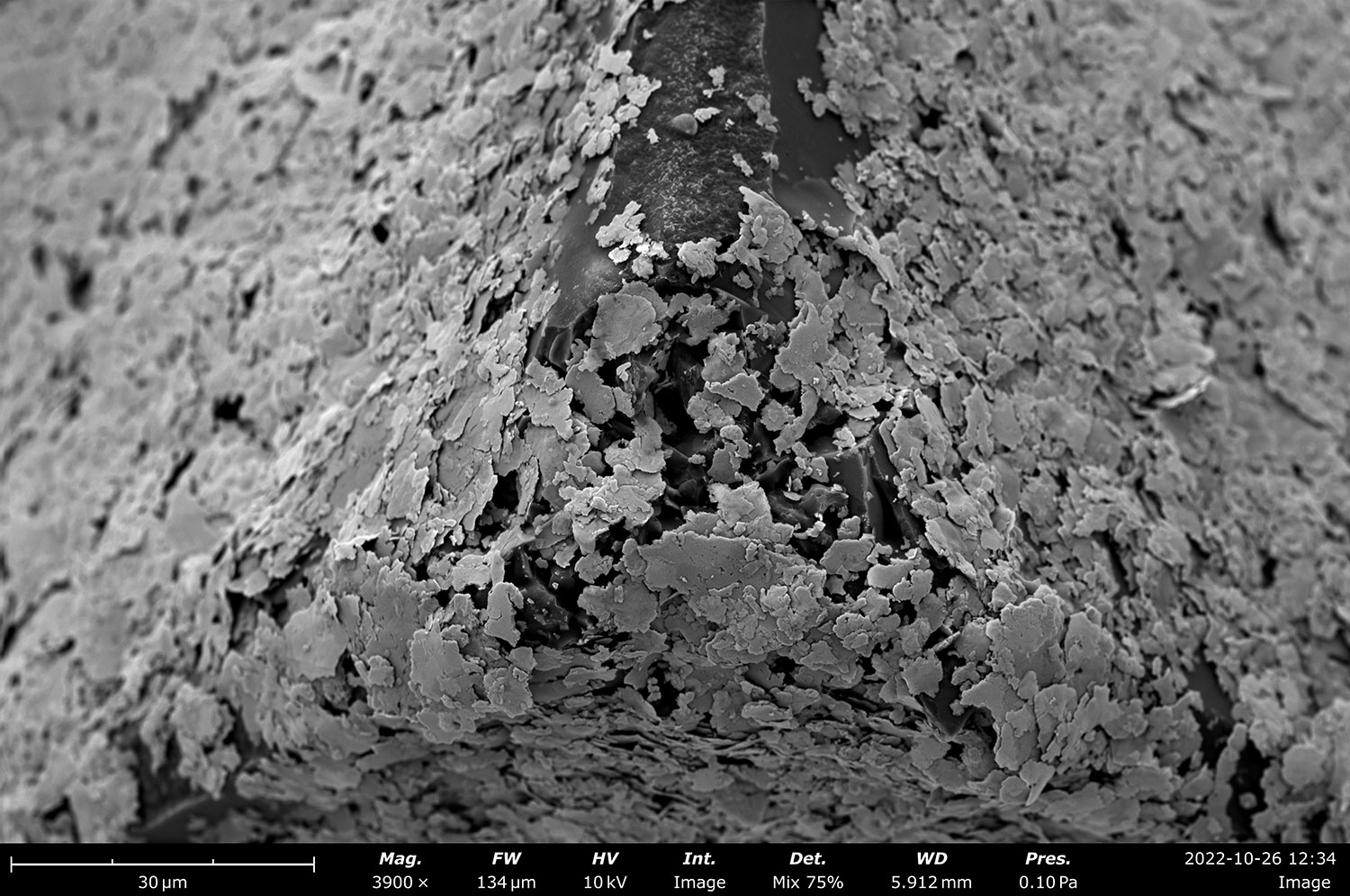
Four-Segment Backscattered Electron Detector (BSD)
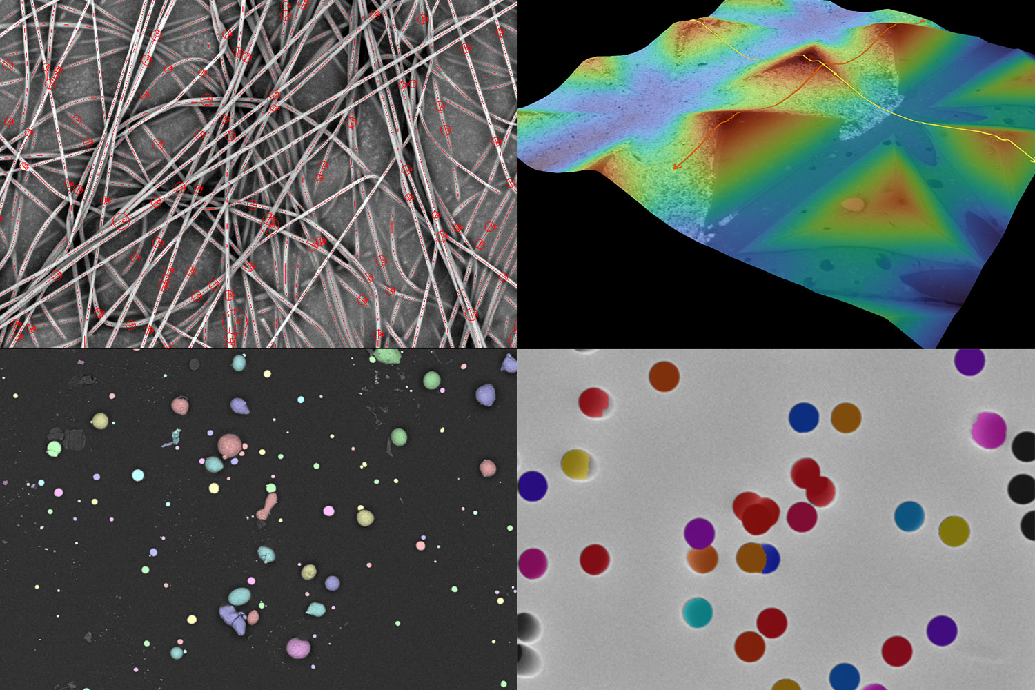
The four-segmented backscattered electron detector (BSD) delivers sharp compositional contrast between light and heavy elements. Included as a standard feature in every Phenom system, it also offers topographic modes for qualitative visualization of surface roughness, enhancing surface detail and material differentiation.
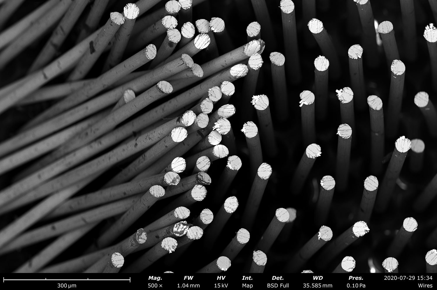
Secondary Electron Detector (SED)
Secondary Electron Detectors (SED) are perfect for high-resolution, surface-sensitive SEM imaging, providing detailed topographical information through secondary electron emissions. This allows for crisp visualization of fine surface features. With an SED, Phenom users can also activate a mixed mode, combining both SED and BSD signals at a customizable ratio for comprehensive imaging analysis.
Energy Dispersive X-ray Spectroscopy Detector (EDS)
Energy Dispersive X-Ray Spectroscopy (EDS) detectors are fully integrated into Phenom SEMs, enabling efficient quantitative surface chemistry analysis. EDS provides rapid and precise results across multiple analysis modes, including regions, points, and lines, making it a versatile tool for a wide range of applications.
Phenom XL Desktop SEM
Accessories
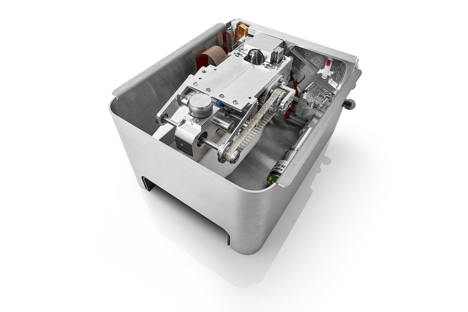
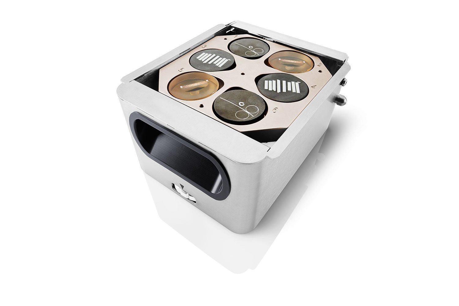
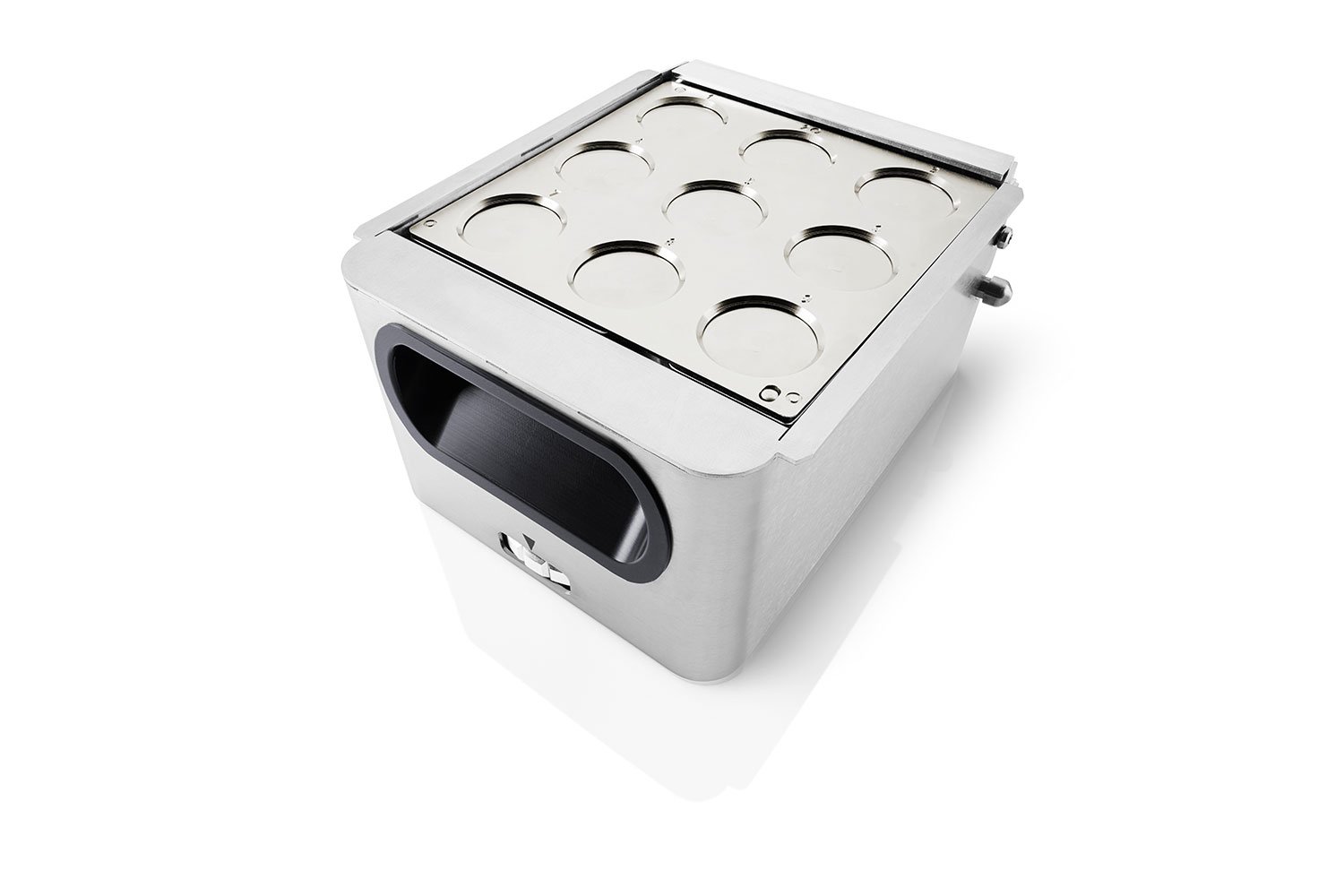
Eucentric Stage
The only eucentric tilt stage available on a desktop SEM, featuring six motorized axes that offer precise control over height, tilt, and rotation, all while maintaining perfect focus on the sample.
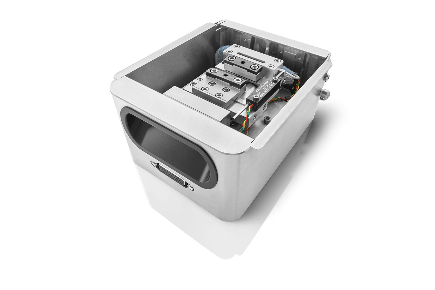
Tensile Testing Stage
Examine material properties in the SEM under forces of up to 1 kN, with the software plotting the force vs. displacement curve.
Resin Mount & Metallurgical Stage Inserts
Ensure a uniform working distance across multiple polished samples, which is especially beneficial for accurate quantitative EDS analysis.
Filter Stage Inserts
Secure delicate filter and membrane samples firmly without the need for carbon tape, ensuring accurate SEM imaging and EDS analysis.
Vacuum Transfer Box
Prepare sensitive samples in a glove box and transfer them in an enclosed transfer box to maintain an inert atmosphere during the transition to the SEM.
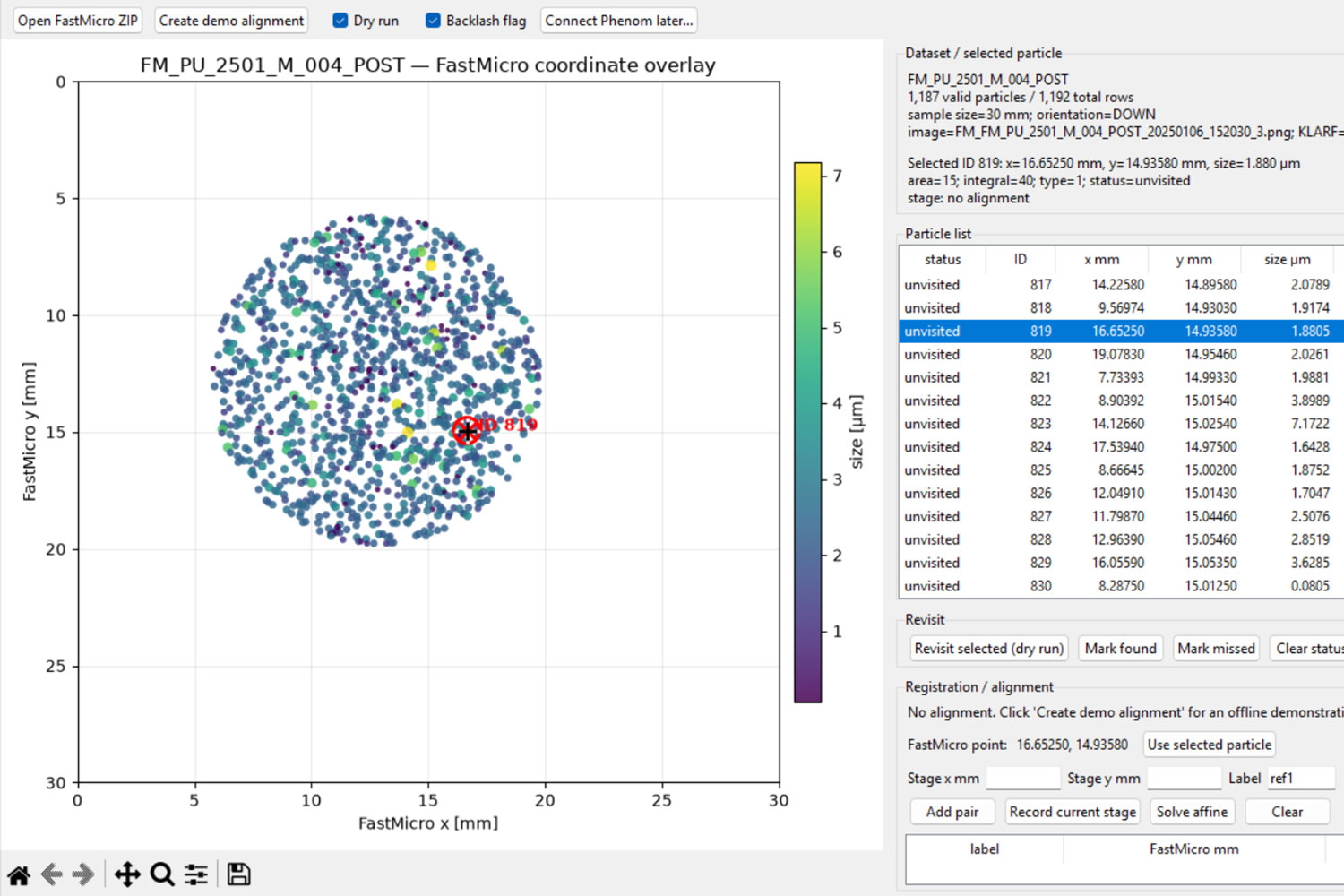
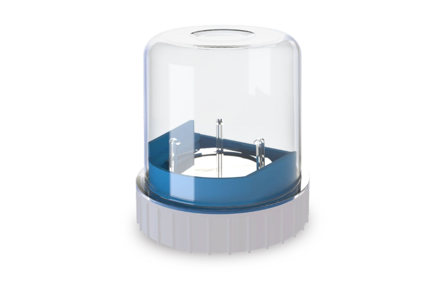
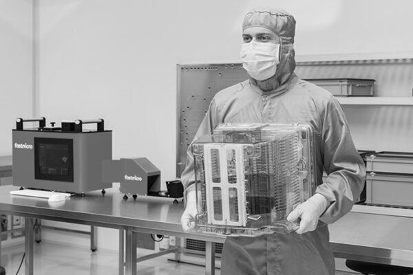
Scattering Particle Analyzer
The Fastmicro Sample Scanner is a compact inspection tool for indirect surface particle contamination measurement, capable of detecting particles down to 0.5 µm using innovative tape-lift samplers. Compatible with a variety of surfaces, including those that are difficult to reach or unusually rough, these samplers leave no measurable residue. With a sample area of 225 mm² analyzed in seconds, the Sample Scanner delivers fast, accurate contamination data across a wide range of industries and applications.
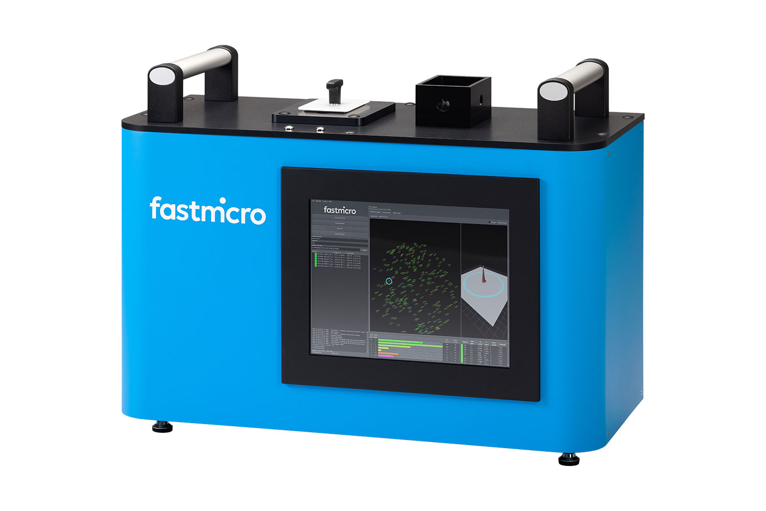
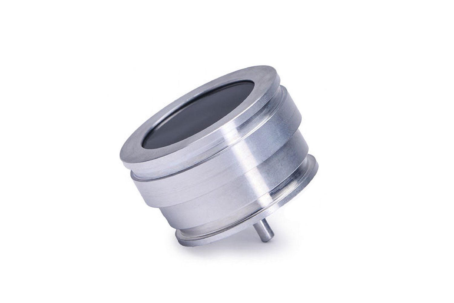
Correlative SEM Data
Particle position data collected by the Sample Scanner can be exported directly for use in correlative scanning electron microscopy (SEM) workflows, enabling automated, targeted analysis of identified particles without the need for manual relocation. To fully enable this workflow, the system can be used with a SEM stub compatible accessory.
Phenom XL Desktop SEM
Software
Python-Based Automation (PPI & PPA)
Expand the capabilities of your Phenom Desktop SEM with PyPhenom, a Python-based library for custom programming. Create your own workflows to control microscope hardware and automate data analysis (PPI) or have us develop a tailored automated workflow for you (PPA).

Phenom Prosuite Software
ProSuite Software, developed specifically for Phenom SEMs, enhances data throughput and eliminates user bias from manual measurements, ensuring more accurate and reliable results. It can extract actionable data on the size and shape of features in SEM images.
ParticleX: Automated SEM/EDS for Particle Analysis
ParticleX is an advanced software package that enables automated data collection, particle analysis, and report generation for Phenom SEM/EDS systems. Ideal for manufacturing environments, it integrates sophisticated algorithms, autonomous workflows, and comprehensive documentation into a single, industry-leading solution.
MAPS
Avizo Trueput
ChemiSEM / ChemiPhase
Streamline elemental and phase analysis with integrated data collection and advanced analysis capabilities. ChemiSEM provides real-time elemental mapping during live imaging and ChemiPhase transforms EDS maps into color-coded phase maps.
Phenom XL Desktop SEM
SEM Sample Preparation
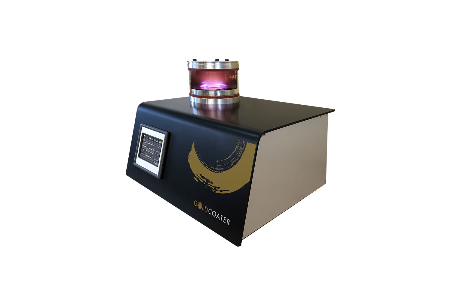
Sputter Coaters
Minimize electric charging and enhance SEM image quality with uniform gold and platinum coatings.
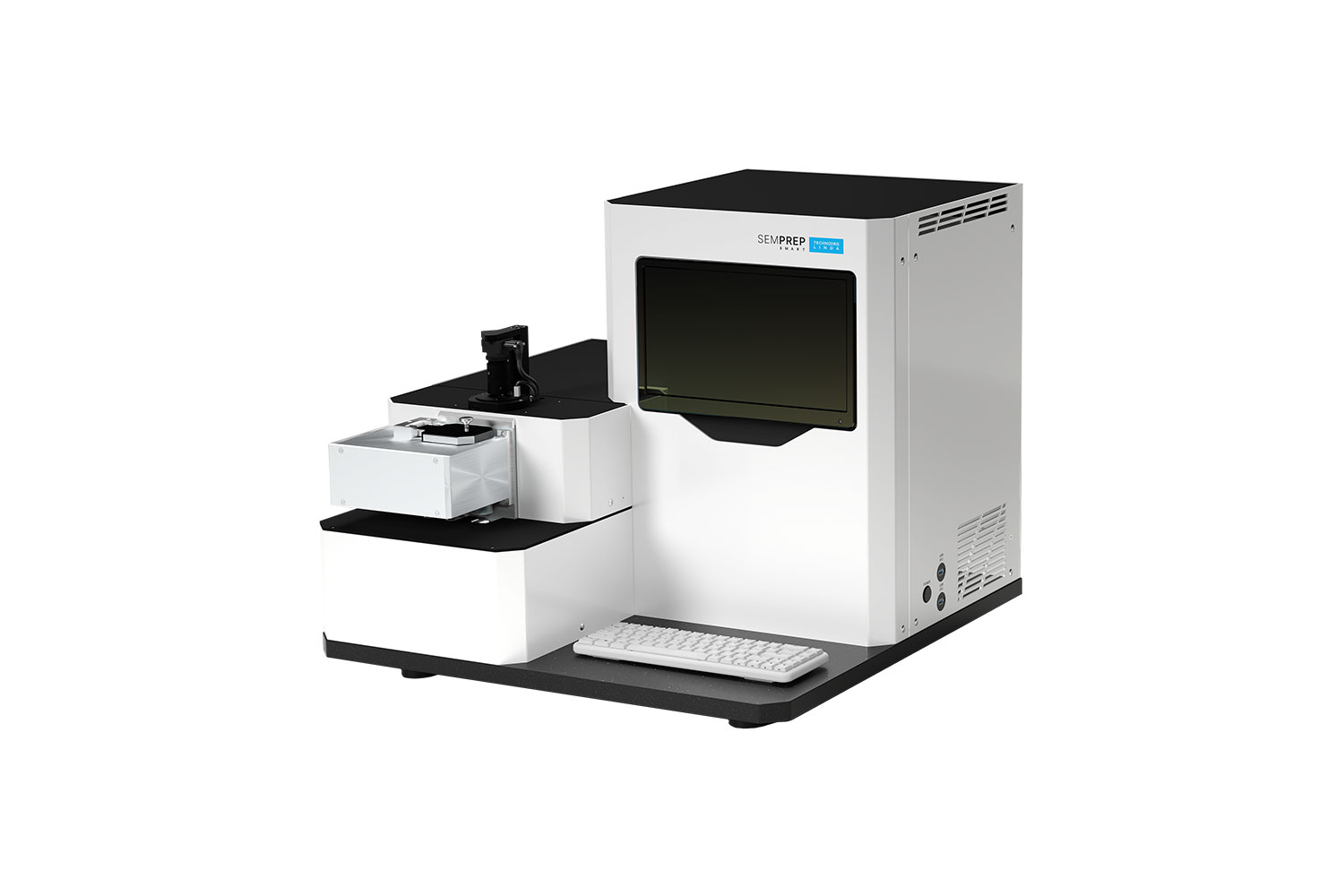
Ion Mills
Prepare artifact-free samples for high quality cross-sectional analysis. Especially useful for EBSD and failure analysis.
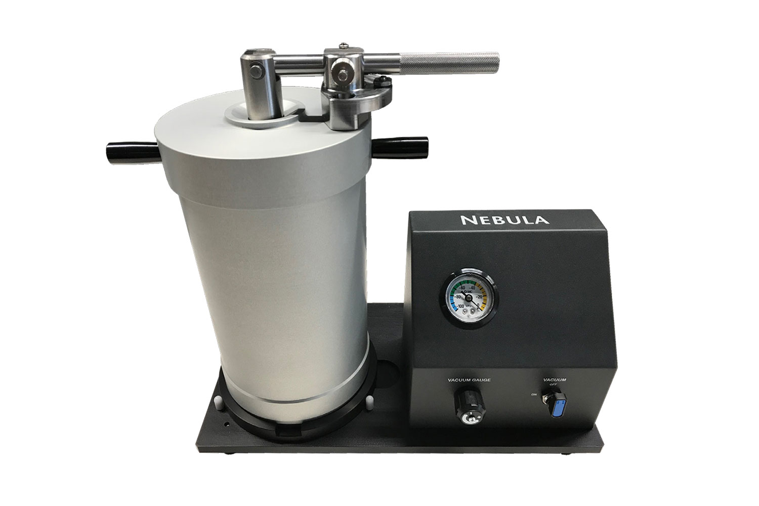
Nebula Particle Disperser
The Nebula is designed to assist in dispersing powder samples, facilitating their preparation for SEM analysis.
Phenom XL Desktop SEM
Product Knowledgebase
Accelerating Surface Particle Contamination Control by Combining Fastmicro and Phenom Technologies
Surface particle contamination remains a critical challenge across advanced manufacturing…
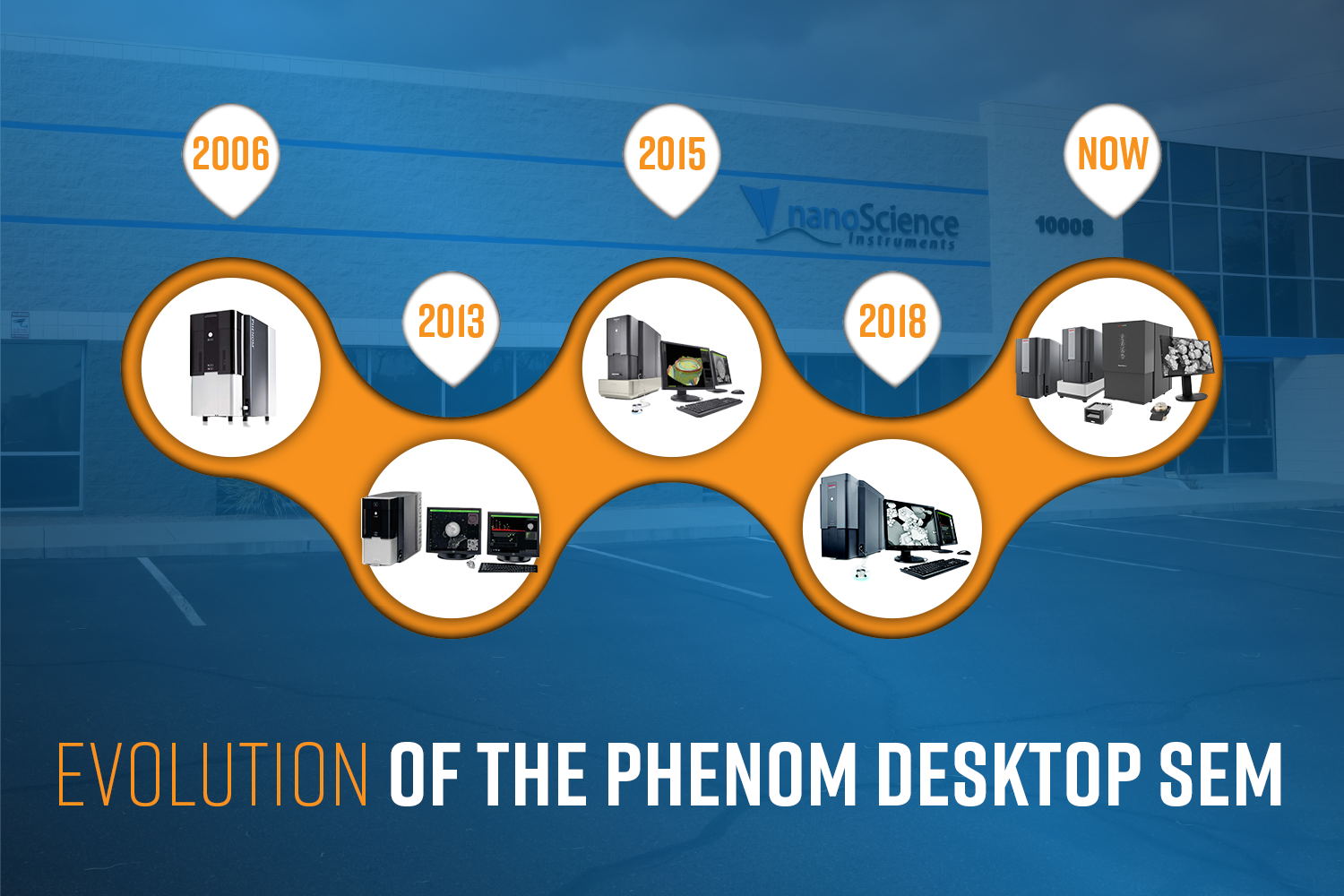
Evolution of the Phenom Desktop SEM
The Phenom Scanning Electron Microscope (SEM) is turning 20 this year! In celebration of t…
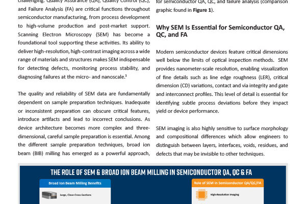
SEM & BIB milling for QA, QC, and Failure Analysis in Semiconductor Devices
Introduction As semiconductor devices continue to scale in complexity and shrink in featur…