PHENOM DESKTOP SEM
Phenom STEM Detector
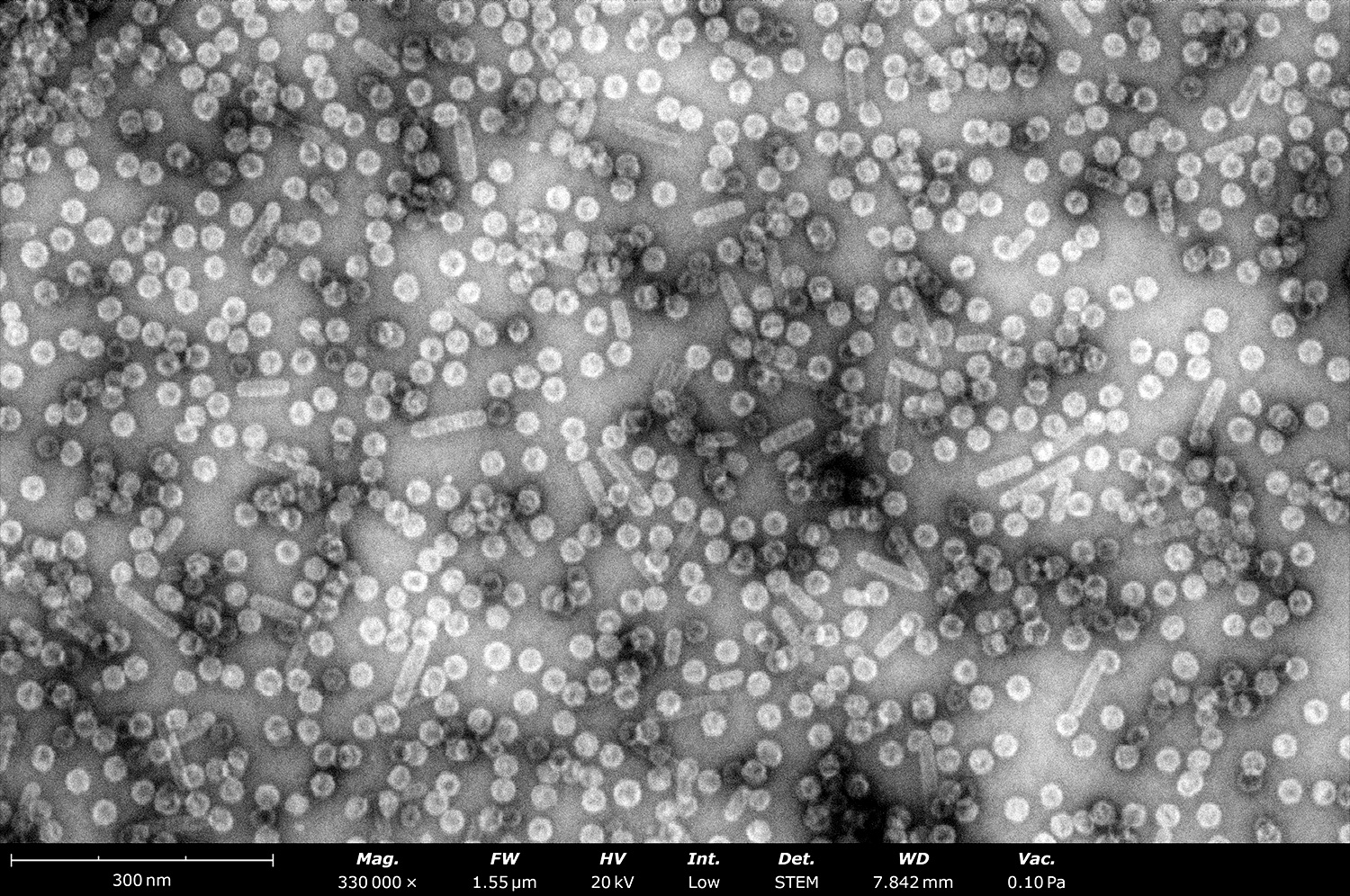
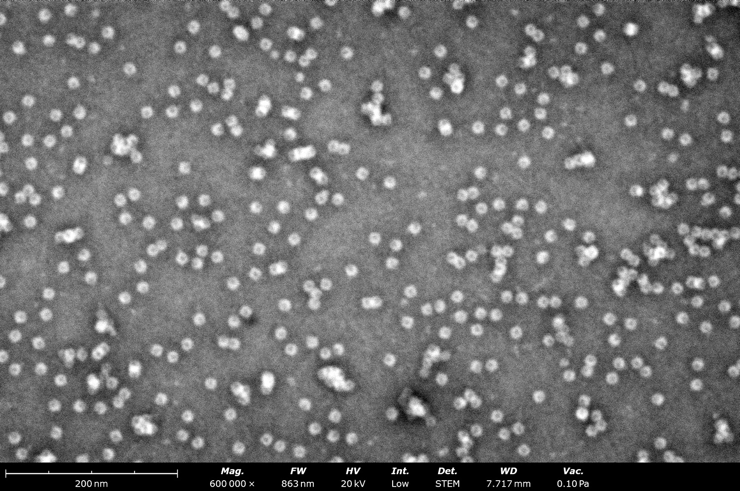
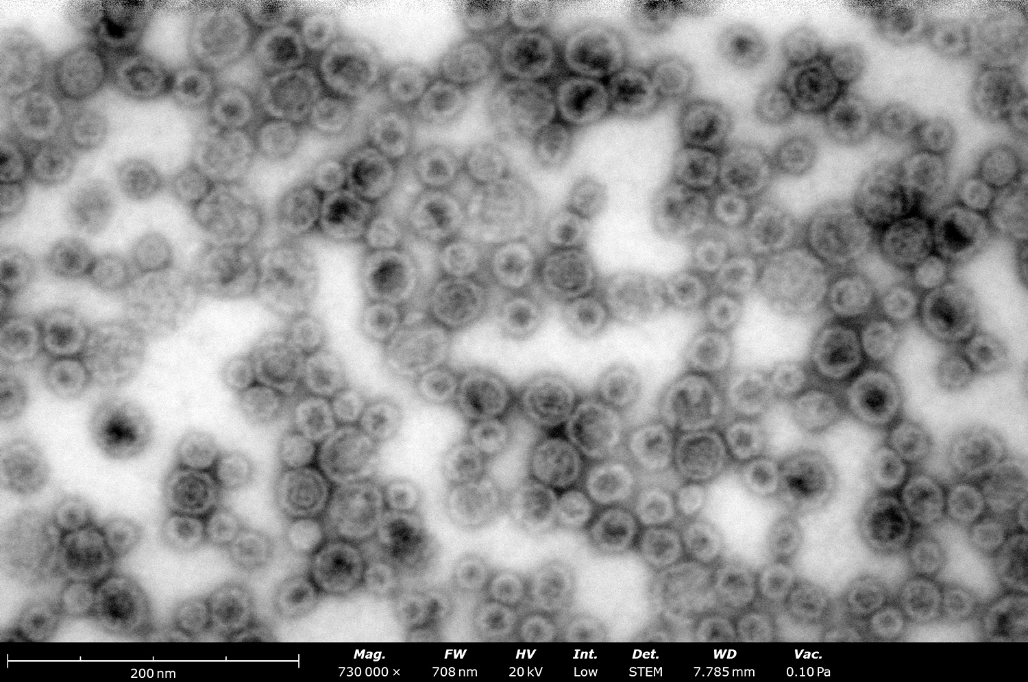
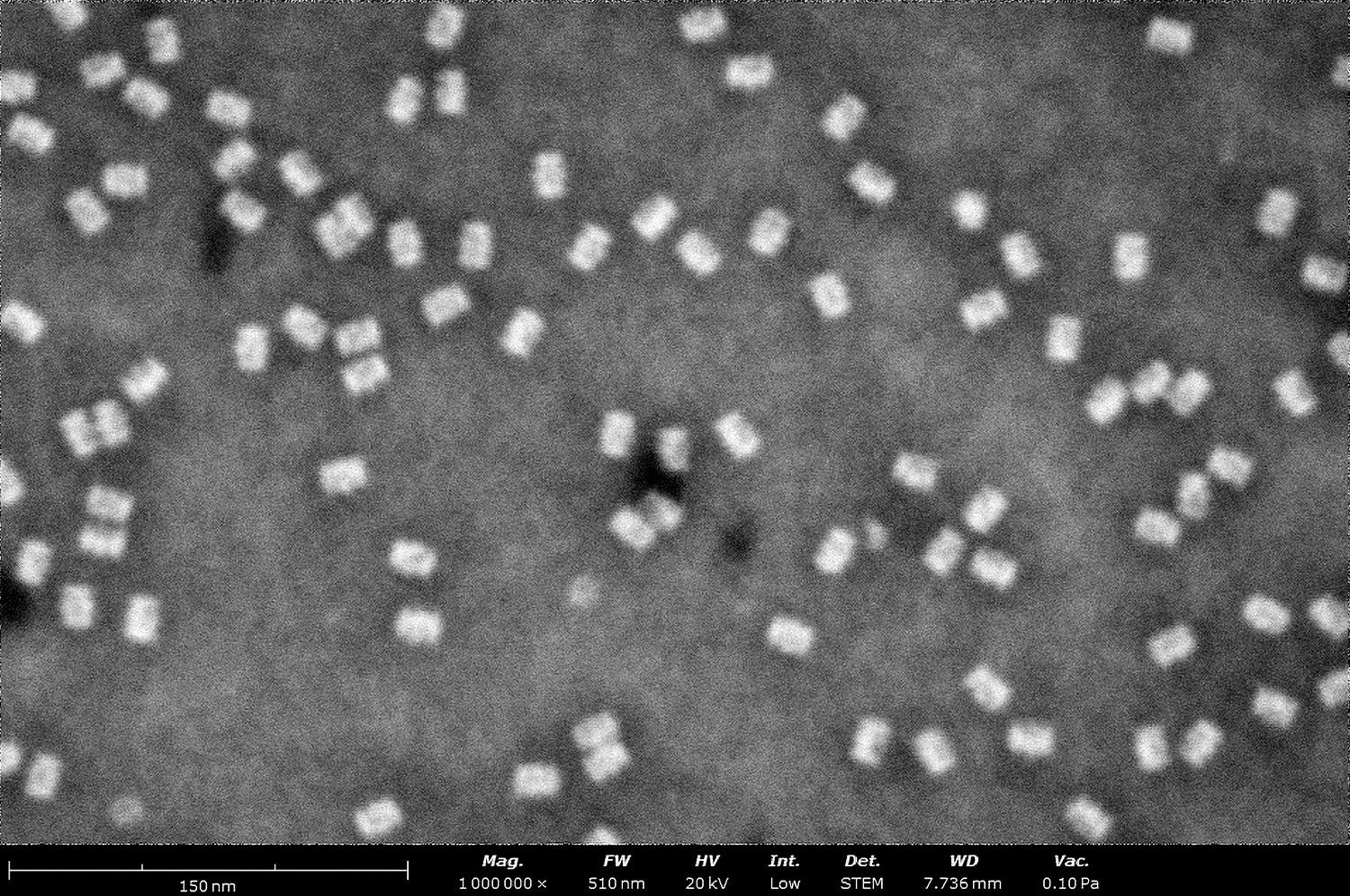
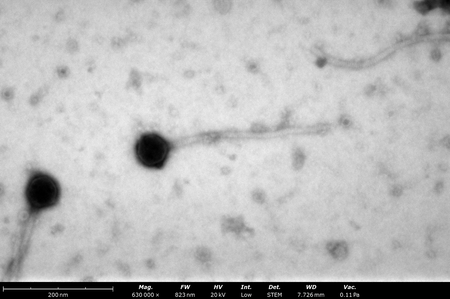
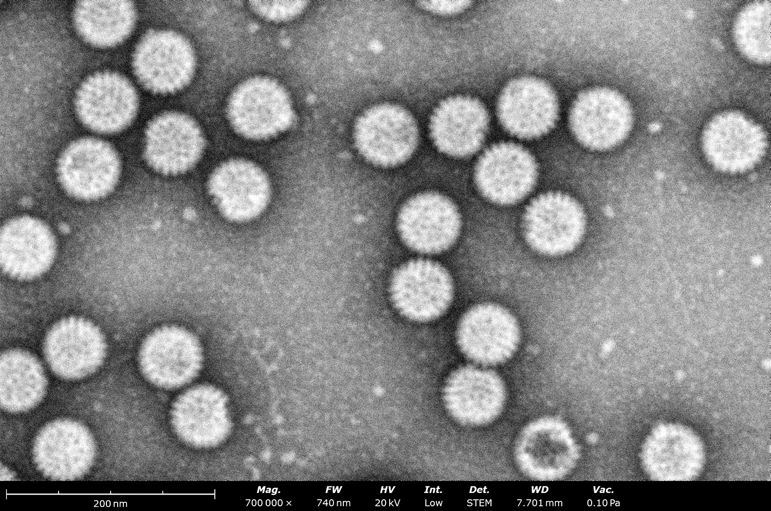
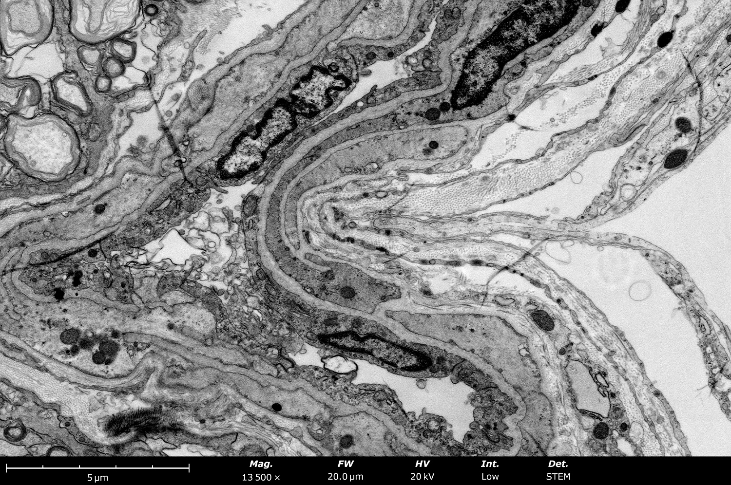
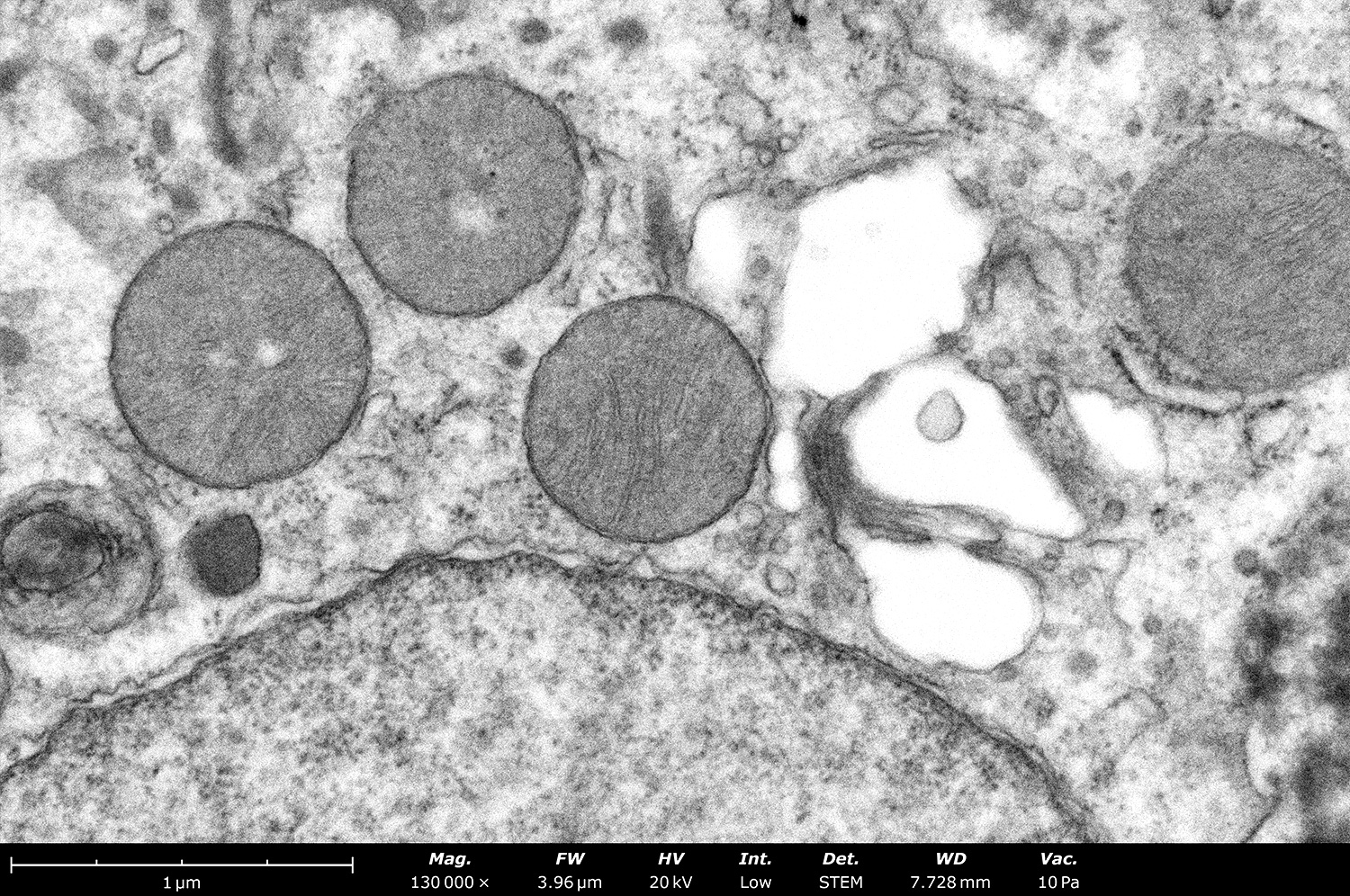
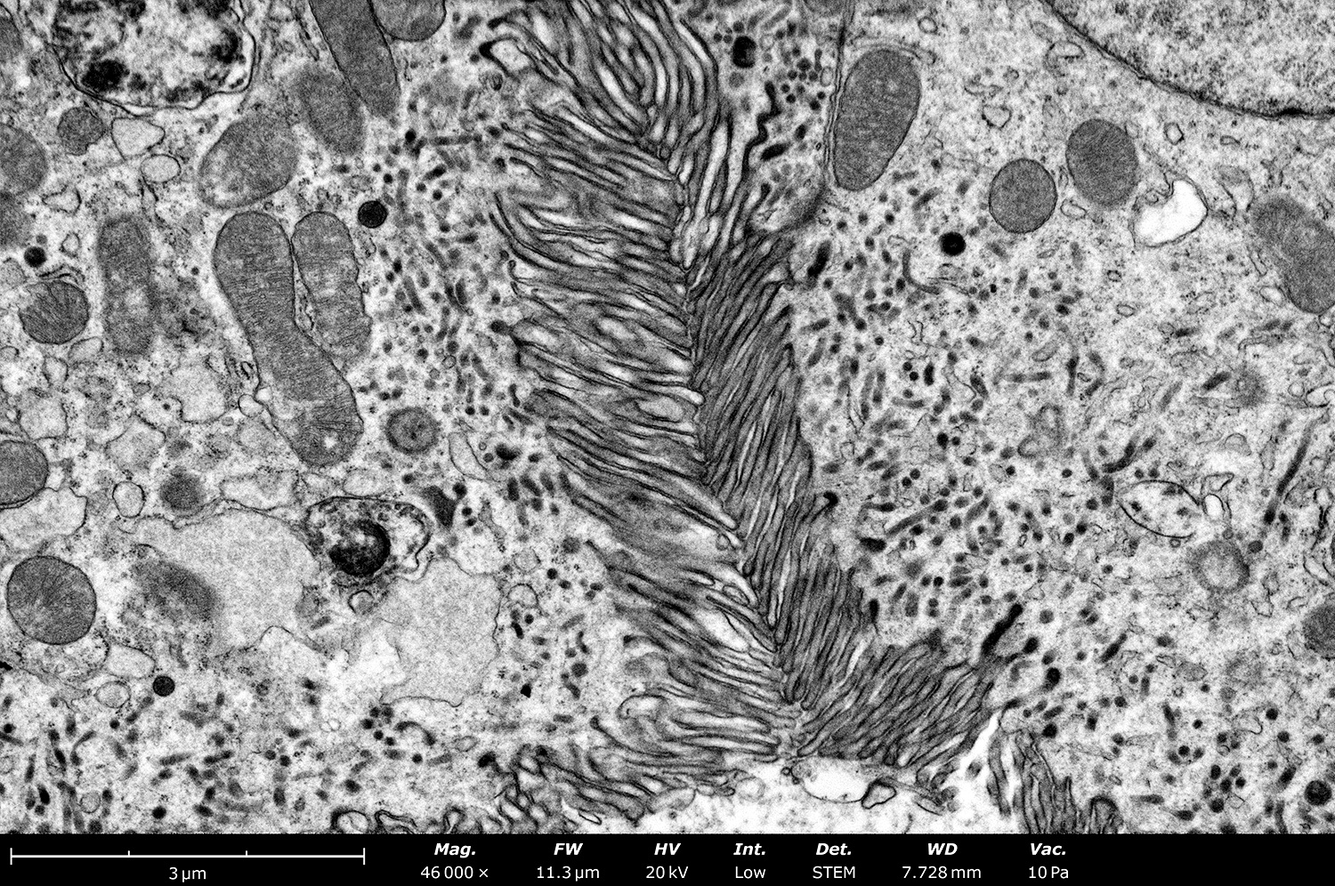
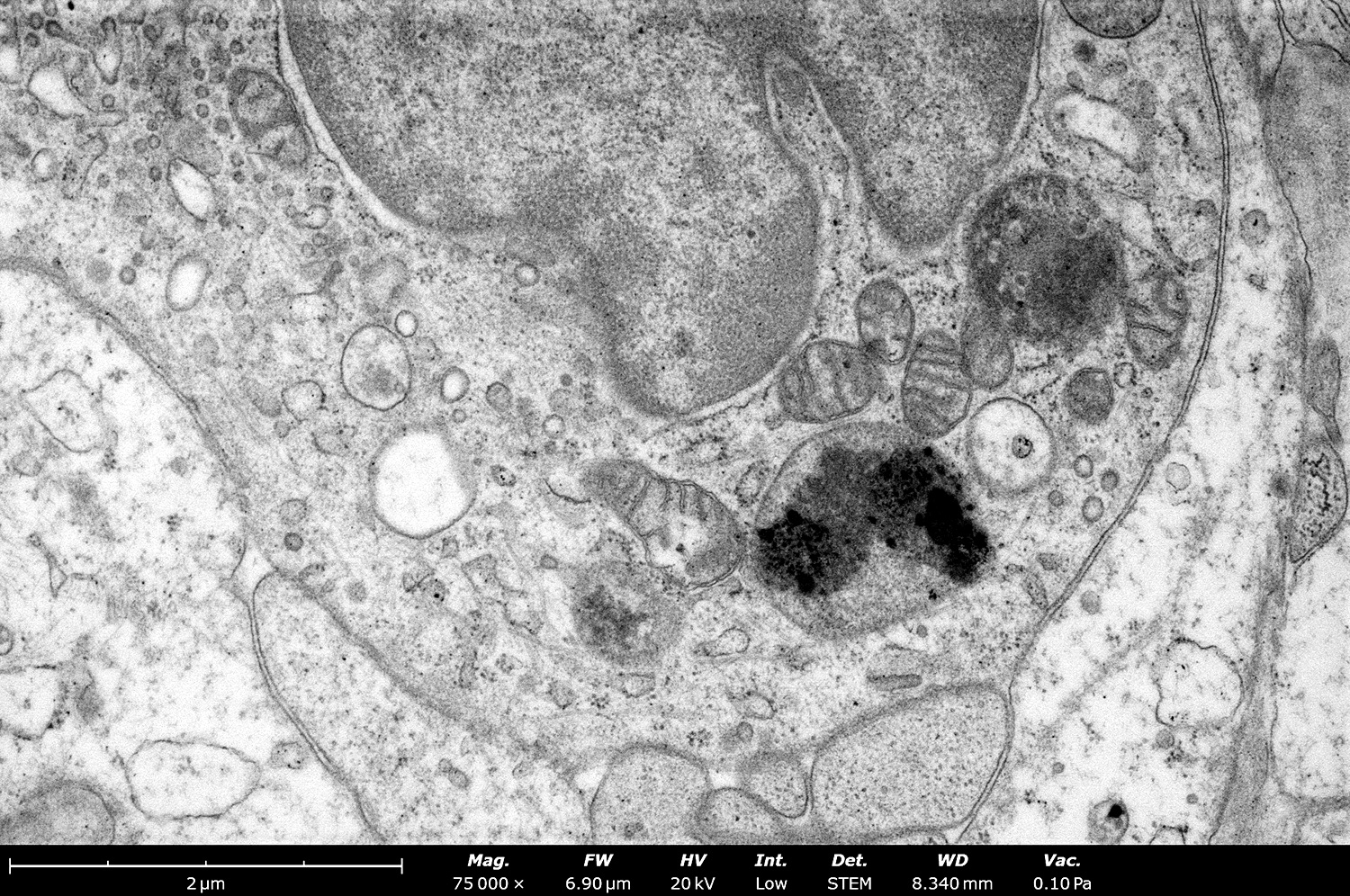
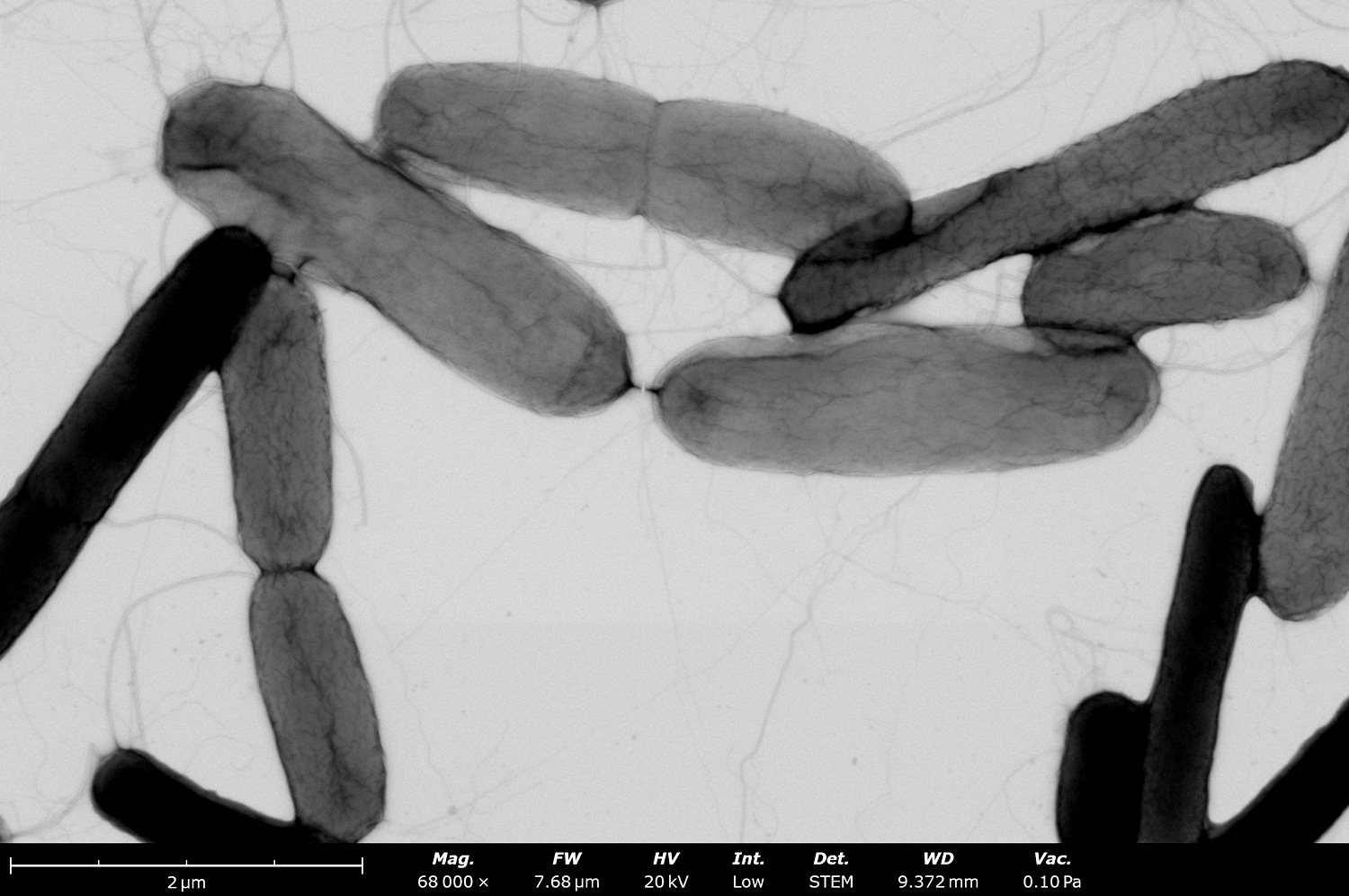
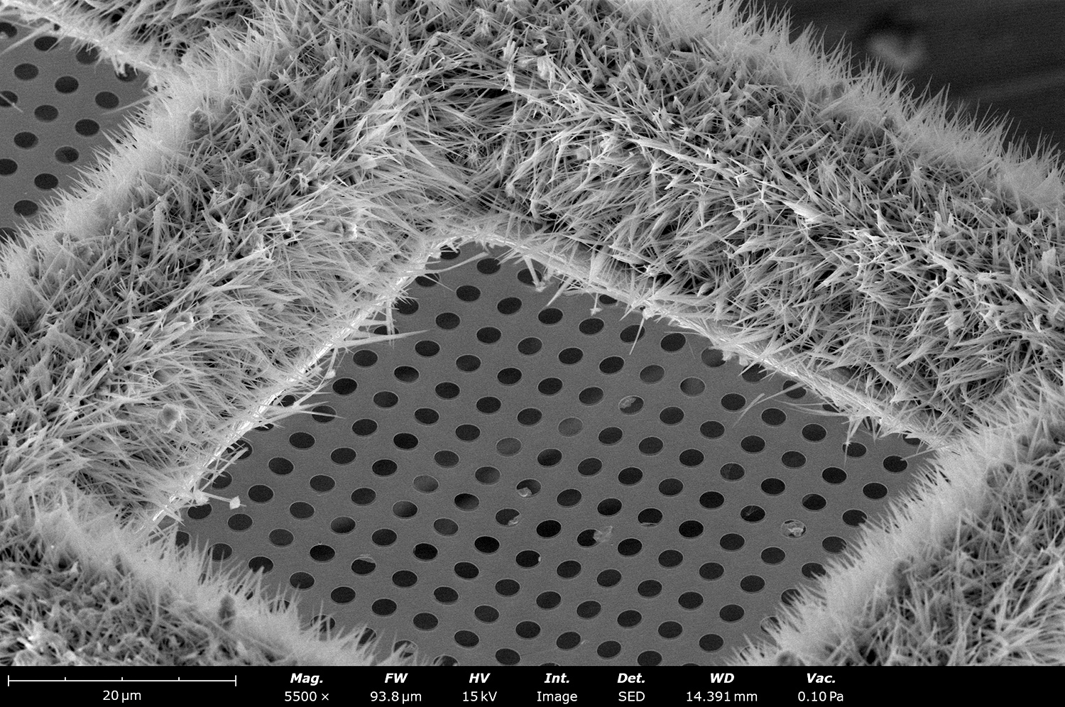
The Phenom STEM (scanning transmission electron microscopy) detector enables transmission imaging for the Phenom Pharos. STEM imaging offers increased visibility of sample morphology and reveals subsurface structural details at higher resolutions than conventional SEM.
STEM Imaging
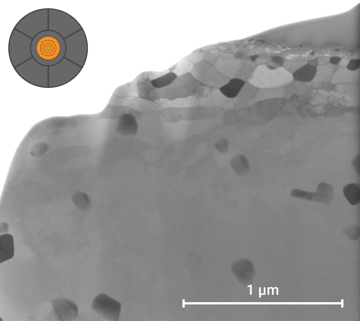
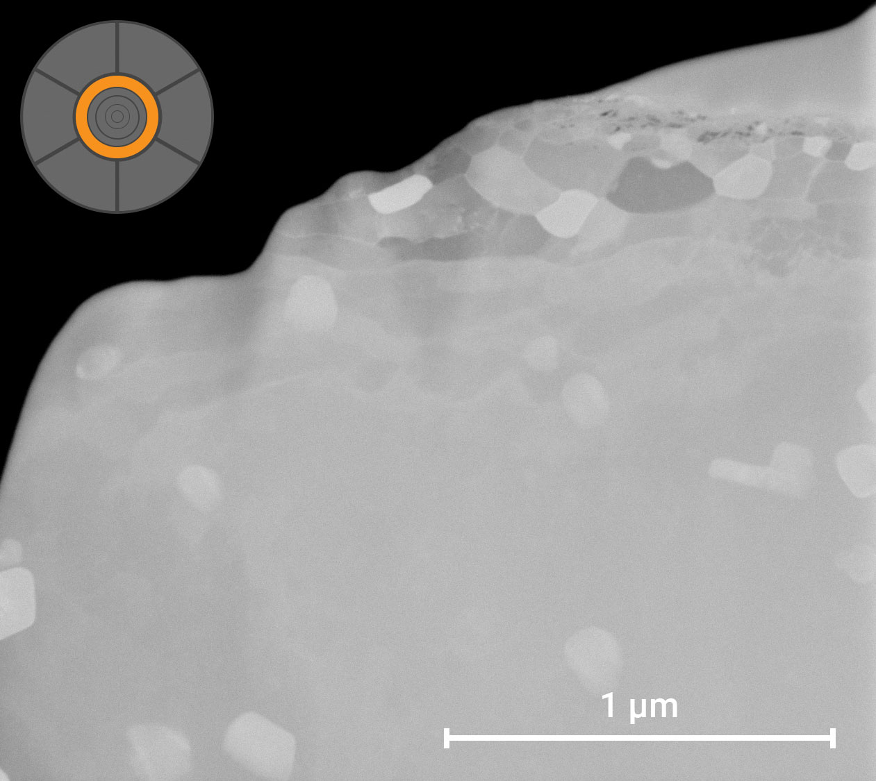
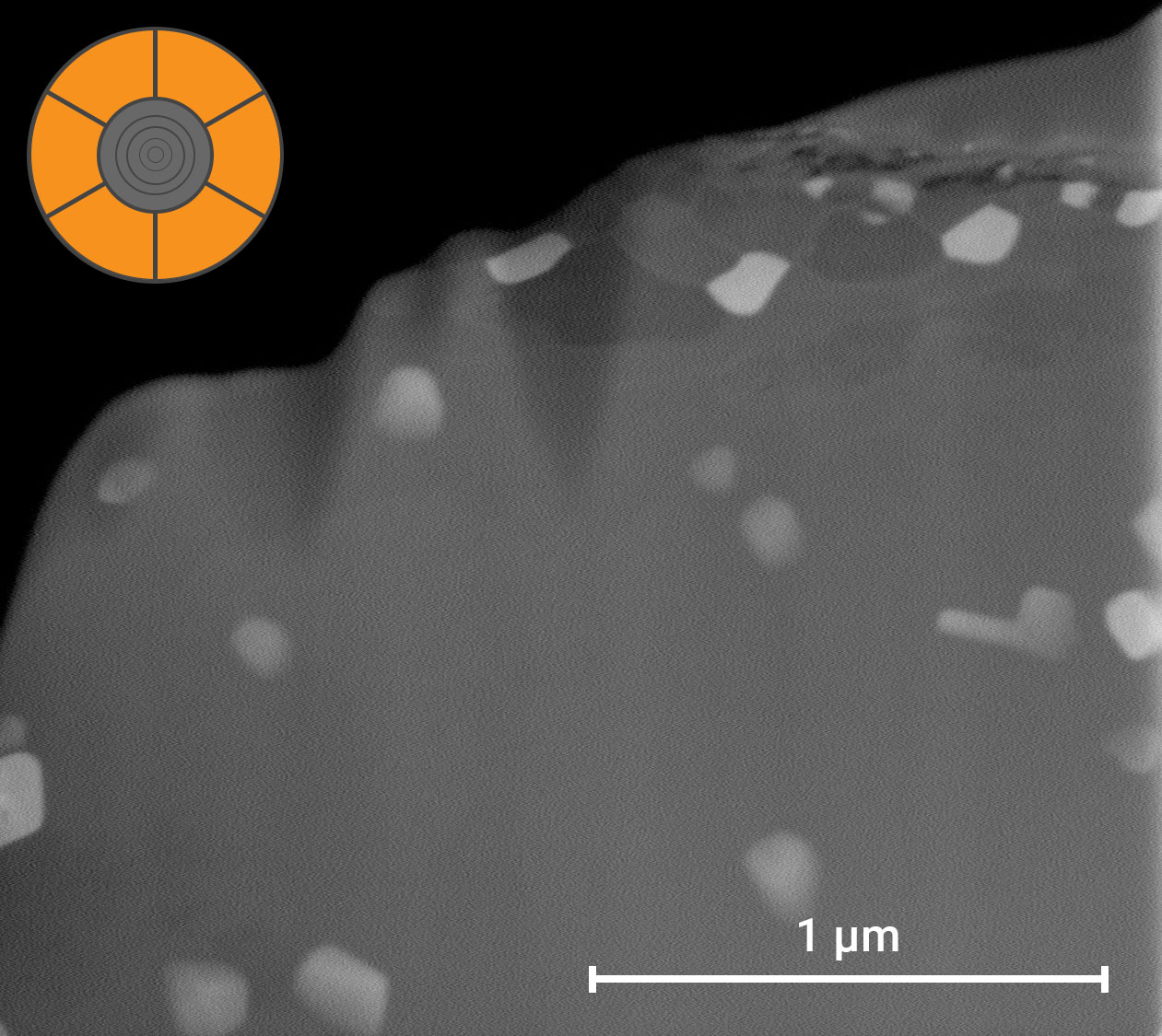
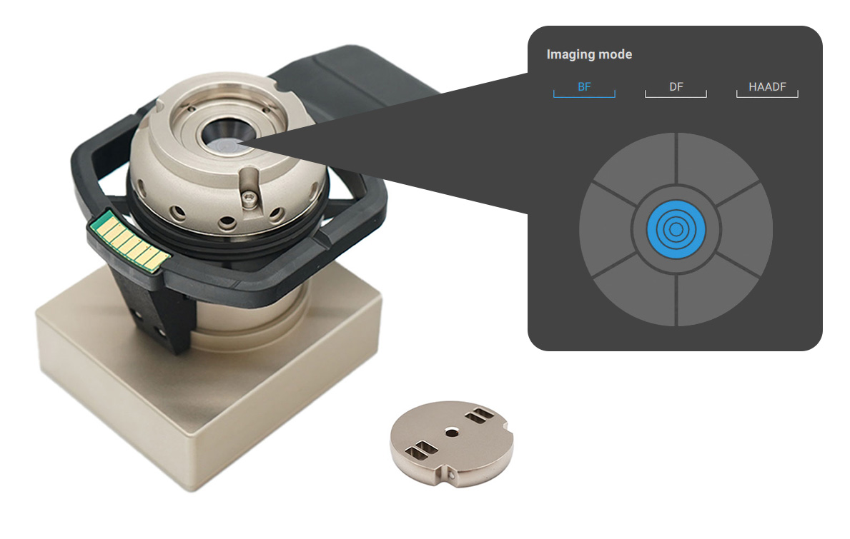
Choose from three standard imaging modes: Bright Field (BF), Dark Field (DF), or High Angle Annular Dark Field (HAADF).
Higher Resolution
With a resolution of <1 nm, STEM achieves a higher resolution than possible with secondary electron detectors.
Application Flexibility
Expand the range of applications accessible to users of any skill level with low-kV STEM imaging on a desktop SEM platform.
Talk to an Instrumentation Specialist Today!
Turnkey Desktop STEM-in-SEM
The small footprint and lower operating vacuum of the Phenom Pharos also provide a faster workflow compared to conventional STEM imaging for applications that do not demand atomic resolution imaging. This new imaging modality provides immense value to a variety of fields including nanomaterials, catalysts, pathology, and batteries.
STEM Imaging Modes:
Bright Field (BF)
BF imaging collects on-axis electrons scattered by the sample. BF image contrast depends primarily on sample thickness and composition, where thicker areas containing heavier elements appear darker. With improved sensitivity to light elements, BF mode can be particularly useful for studying organic samples.
Dark Field (DF)
DF imaging detects off-axis electrons that result from relatively lower diffraction angles. Image contrast depends on thickness and atomic number with brighter areas corresponding to local mass-thickness. DF imaging is more sensitive to atomic number differences in lighter elements, and is useful for a broad range of samples.
High Angle Annular Dark Field (HAADF)
HAADF imaging collects the off-axis signals at the highest scattering angles and is most sensitive to atomic number contrast, or Z contrast. HAADF is particularly sensitive to heavier elements such as metal atoms. This mode can be used to detect features that are harder to visualize with the other imaging modes.
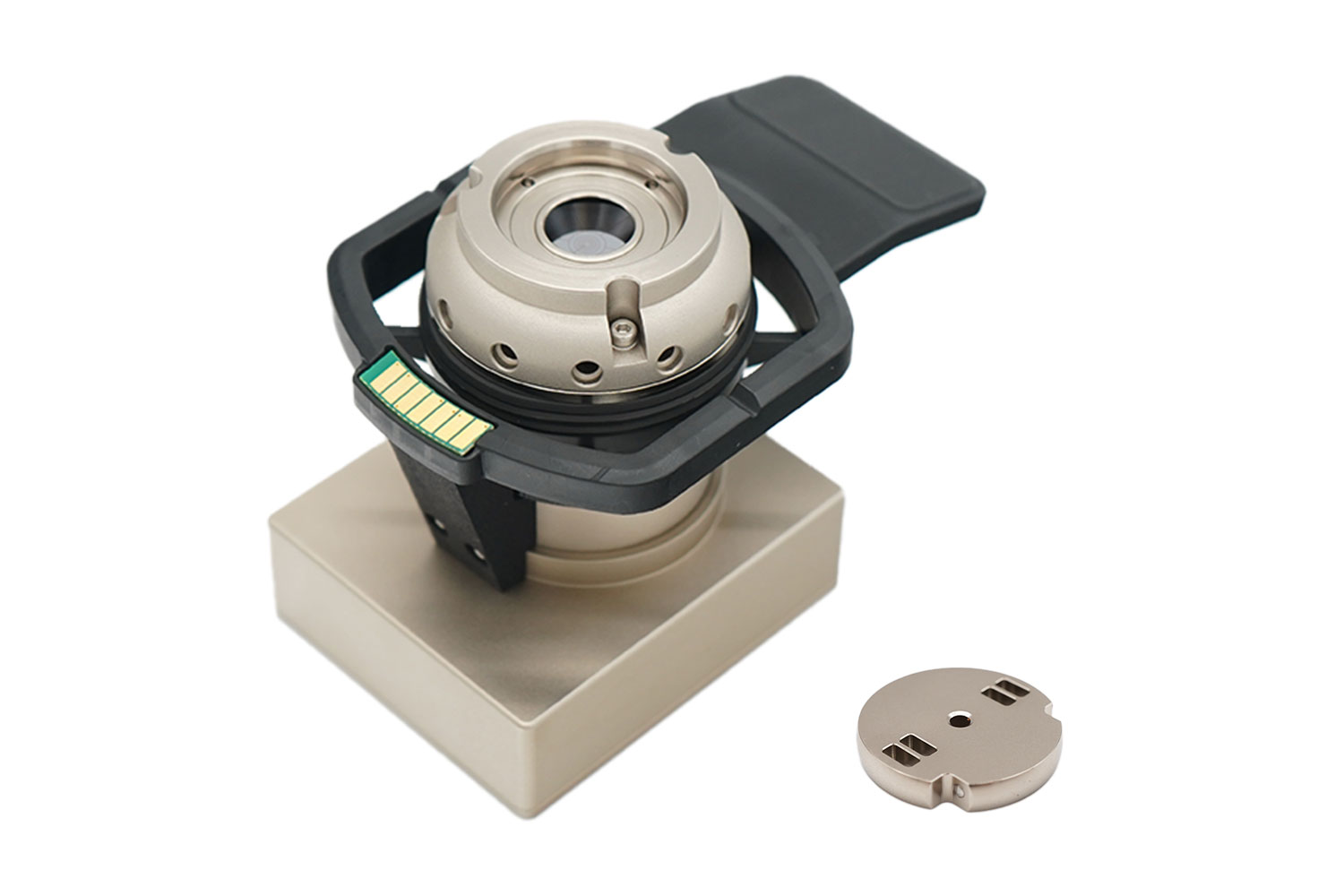
STEM Detector for Phenom Pharos
Product Features
Integrated STEM Detector and Sample Holder
The sample holder features an integrated detector with 11 segments, offering presets for bright field (BF), dark field (DF), and high-angle annular dark field (HAADF), along with options for custom configurations. Its camera length is optimized for a wide range of applications, including life sciences. This smart design removes the need for cables, as the interface board automatically detects the STEM detector and adjusts to the available imaging conditions.
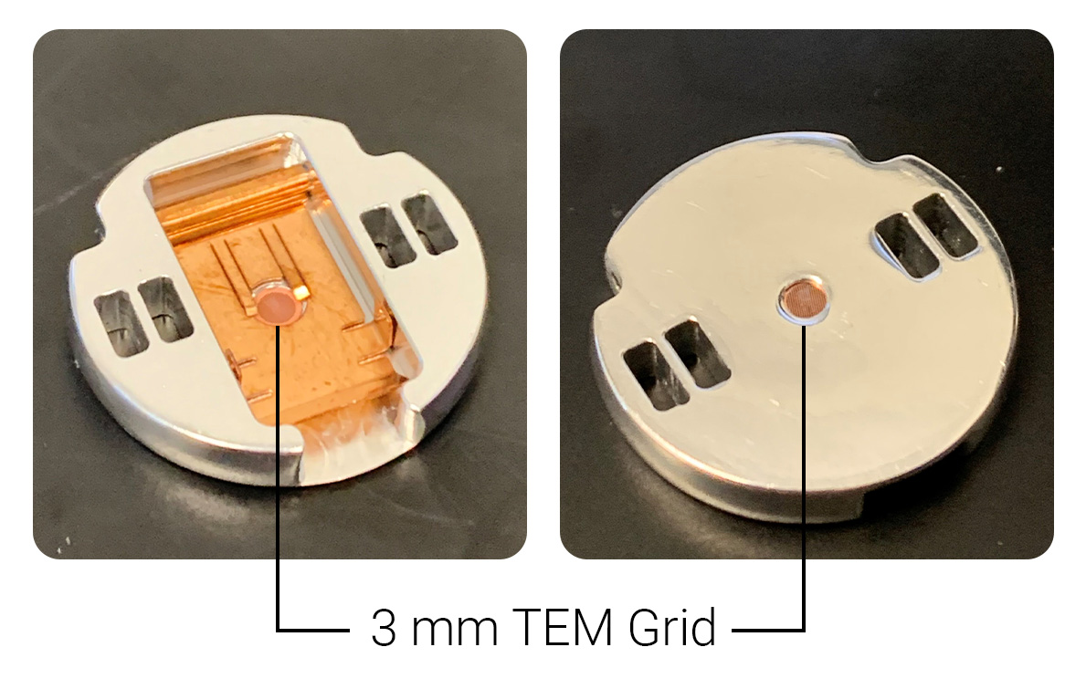
Easy sample loading
The sample chuck securely holds standard 3-mm TEM grids, which can be loaded into the detector in either orientation. Built for durability, the sample mount is designed to handle frequent sample exchanges and repeated loading and unloading cycles. The stub is optimized with a copper clamp that holds the grids in place, guaranteeing sample flatness and grid protection.
Time to image <40 seconds
After mounting the sample in the stub, the Phenom Desktop STEM fast pump down mechanism and user interface makes it possible to begin imaging in less than one minute. The UI automatically recognizes the STEM detector and instantaneously enables the STEM imaging pane. After selecting your preferred imaging conditions (BF, DF, or HAADF), the detector is read out accordingly and area of interest is imaged with the optimal signal-to-noise ratio.
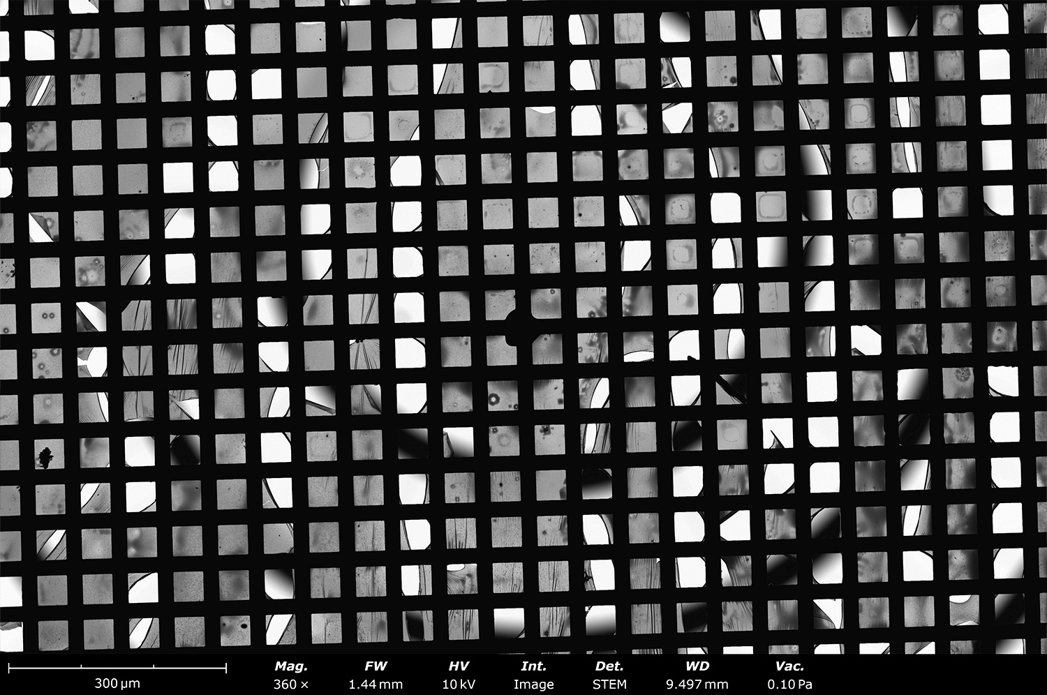
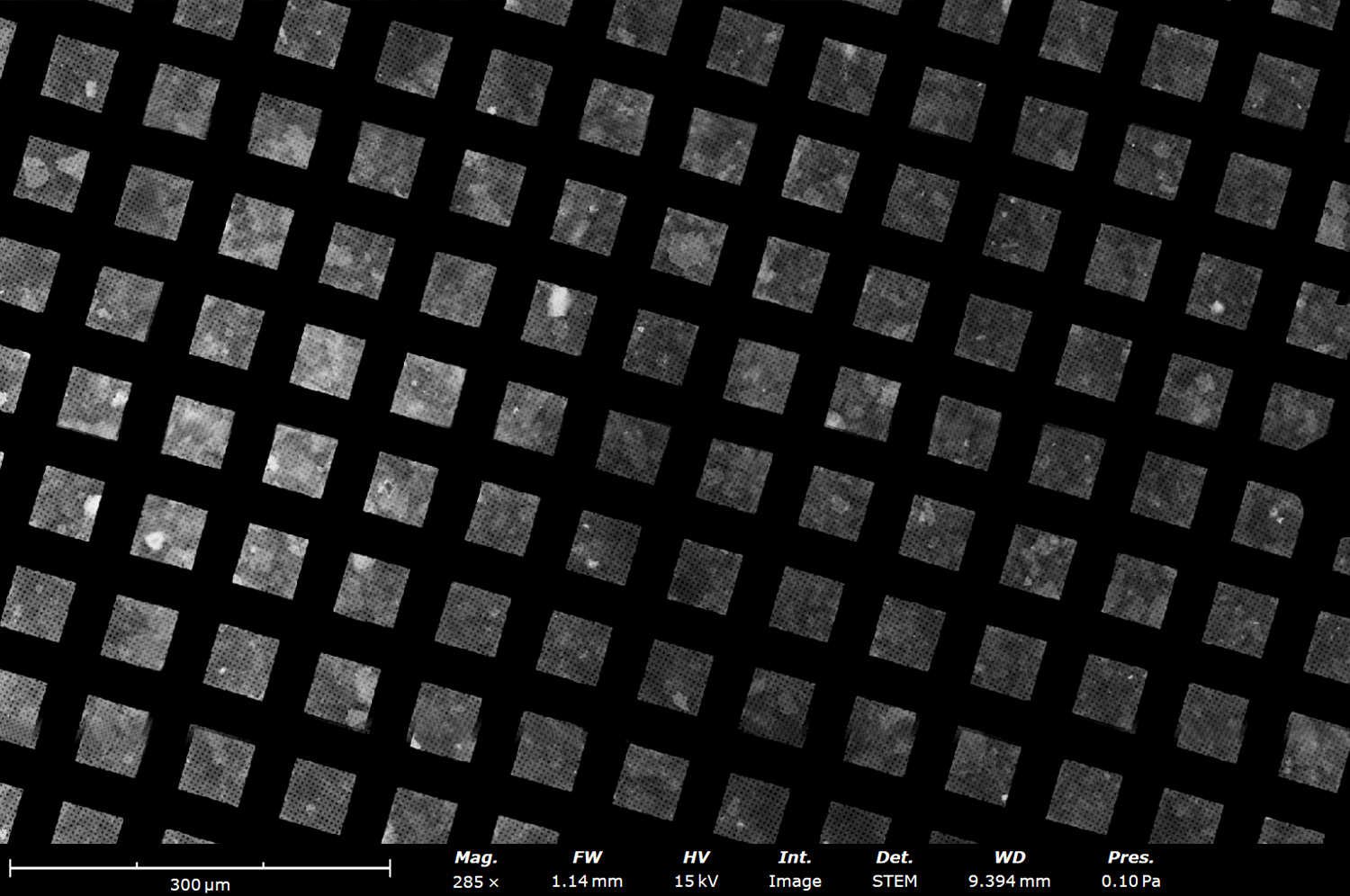
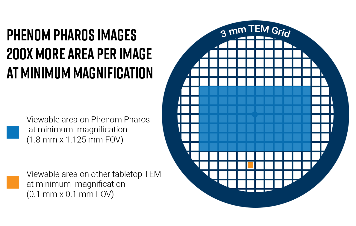
Expansive imaging area
With the large pixel array detector, you can view nearly the entire sample grid at minimum magnification. This advanced feature offers 200 times more field of view compared to other TEM systems, making it a great solution for quickly screening grid quality and identifying areas of interest.
STEM Detector for Phenom Pharos
Product Knowledgebase
Tissue Section Analysis with Benchtop STEM
Furthering our collective understanding of biological processes is a complex, demanding en…
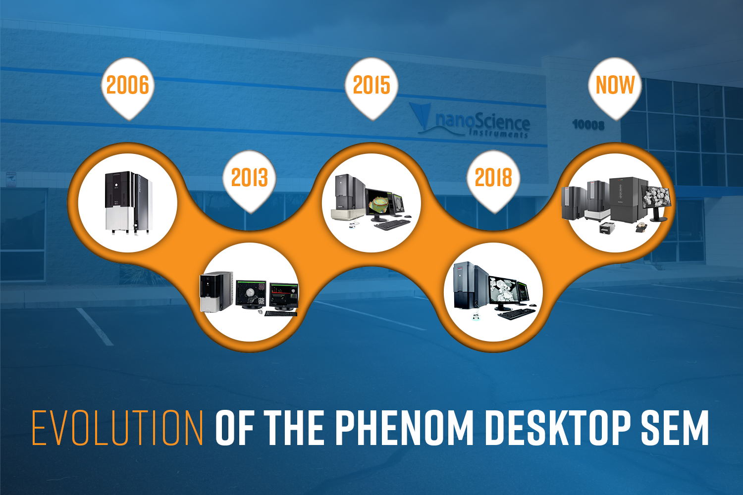
Evolution of the Phenom Desktop SEM
The Phenom Scanning Electron Microscope (SEM) is turning 20 this year! In celebration of t…
Phenom Pharos: A Compact Desktop STEM for Screening Negative Stained Samples
The Phenom Pharos Desktop SEM/STEM is a compact and affordable solution designed for cryo-…