The Centre for Ultrastructural Imaging (“CUI”) is the central electron microscopy unit at King’s College London, one of the most advanced EM hubs in Europe.
Professor Roland Fleck and his team are using SenseAI software to apply new Volume Electron Microscopy (vEM) imaging workflows, resulting in a 4x increase in acquisition speed with enhanced image quality.
The SenseAI software integrates with the CUI’s existing electron microscopy equipment to overcome existing vEM limitations such as:
- Complex Materials: Materials are complicated and difficult to image using an electron microscope. Imaging and analysis of these materials takes great care and accuracy.
- Acquisition Time: Conventional volume imaging techniques are time-consuming, limiting the pace of research.
- Beam Damage: All electron microscopy suffers from beam damage, but this effect is predominantly limiting for the imaging of cryogenically frozen biological materials; the materials are very sensitive and also likely to be melted.
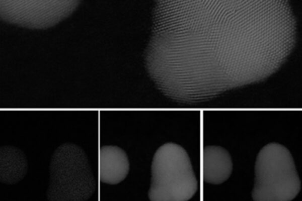
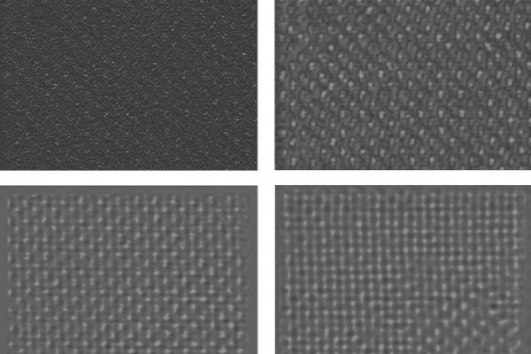
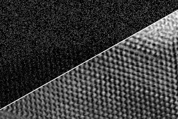
Specifically, a new paper by King’s College, SenseAI, JEOL and others, accepted for publication to the Journal of Microscopy, highlights a 4x improvement in imaging time i.e. acquiring only 25% of the pixels using the SenseAI compressed sensing technique took only 25% of the time compared to the 100% sampled data. Crucially, reconstructed images retained high-frequency detail and, in many cases, showed improved contrast and interpretability compared to conventional full-dose raster scans.
In this webinar, you get an introduction to the SenseAI technology and have the chance to interact with Dr. Nigel Browning, the inventor of SenseAI, and Dr. Roland Fleck. Bring along your questions, pain points, and experiences as we open up the floor on the topic of Volume Imaging!