Electrospun Scaffolding for Cell Growth and Tissue Engineering
In any type of scaffolding material cell migration, proliferation and differentiation are a key parameter for the success of tissue engineering and regenerative medicine applications. The ability of electrospun scaffolds to biomimic the extracellular matrix (ECM) environment allows the electrospinning technique to be an ideal choice for tissue engineering and regenerative medicine. Typical electrospun scaffolds are created with aligned or random fiber orientations that mimic different tissue environment. Researchers have exploited this fiber orientation feature to explore therapeutic strategies by studying the ability of cells to grow in vitro in different environments to mimic the in vivo counterpart.
Nanofiber Alignment for Medical Grade Application
Various ways to create aligned and random fibers have been studied over the past few decades. Among the most typical are the use of rotating drums at different linear speeds. While random fibers can be obtained at low rpm’s, aligned fibers are able to be created at high linear speeds. The Fluidnatek® line allows the process of aligned and random fibers using a rotating drum, disk, and mandrel which all can be used up to 2,000 rpms and 30 cm in diameter. The electrospinning equipment can be used with any natural and synthetic polymers, along with their blended forms, to create different fiber orientation. The cabin of the units are chemically resistant and all include a solvent exhaust port, these two features that are critical when strong solvents, like acids and organics, are needed in solution to create tissue-engineered scaffolds.
Advantages of Fiber Orientation for Tissue Engineering
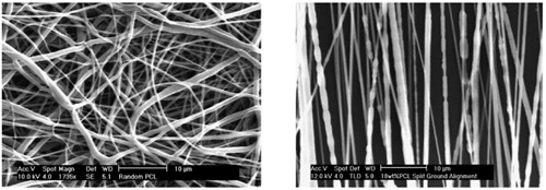
The scanning electron microscope (SEM) images below show random and aligned polycaprolactone (PCL) fibers made with the Fluidnatek® electrospinning equipment. Cell seeding onto these oriented electrospun fibers can be implemented in vitro to study the behavioral growth of different types of cells before developing the appropriate scaffolding material for tissue engineering applications. The use of bioactives into these fibers can also be implemented with the Fluidnatek®equipment to properly engineer a scaffold to target cell growth and regenerative medicine.
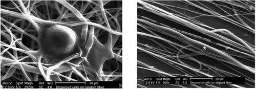
Cell migration can be inhibited by random electrospun PCL fibers, or the commonly used solid polystyrene cell culture dish, where the use of aligned PCL fibers can render realistic in vitro results similar to the ones reported in vivo. As an example, the migration of glioma stem-cell demonstrated to have average speeds of 0.8 ± 0.08 µm h-1 on random fibers and 4.2 ± 0.39 µm h-1 on aligned fibers. These results are due to the randomly oriented substrate hindering cell proliferation in random fibers. However, when aligned fibers are employed cell proliferation is higher and faster. Better pharmacological strategies to treat and inhibit specific types of cancers can be implemented by studying cell proliferation with these types of fibers. The following video shows how glioma cells grow and propagate in aligned PCL fibers over time.
Time lapse of glioma cells growing and propagating in aligned electrospun PCL fibers