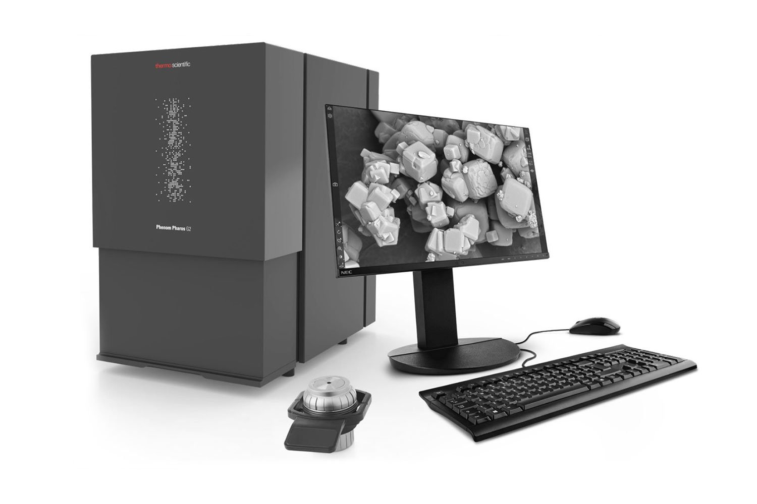
Phenom Desktop SEMs, also known as benchtop or tabletop SEMs, embody the functionality and capabilities of larger, floor-standing SEM models, while introducing specialized software analysis tools and a level of versatility of their own that caters to the needs of modern laboratory settings.
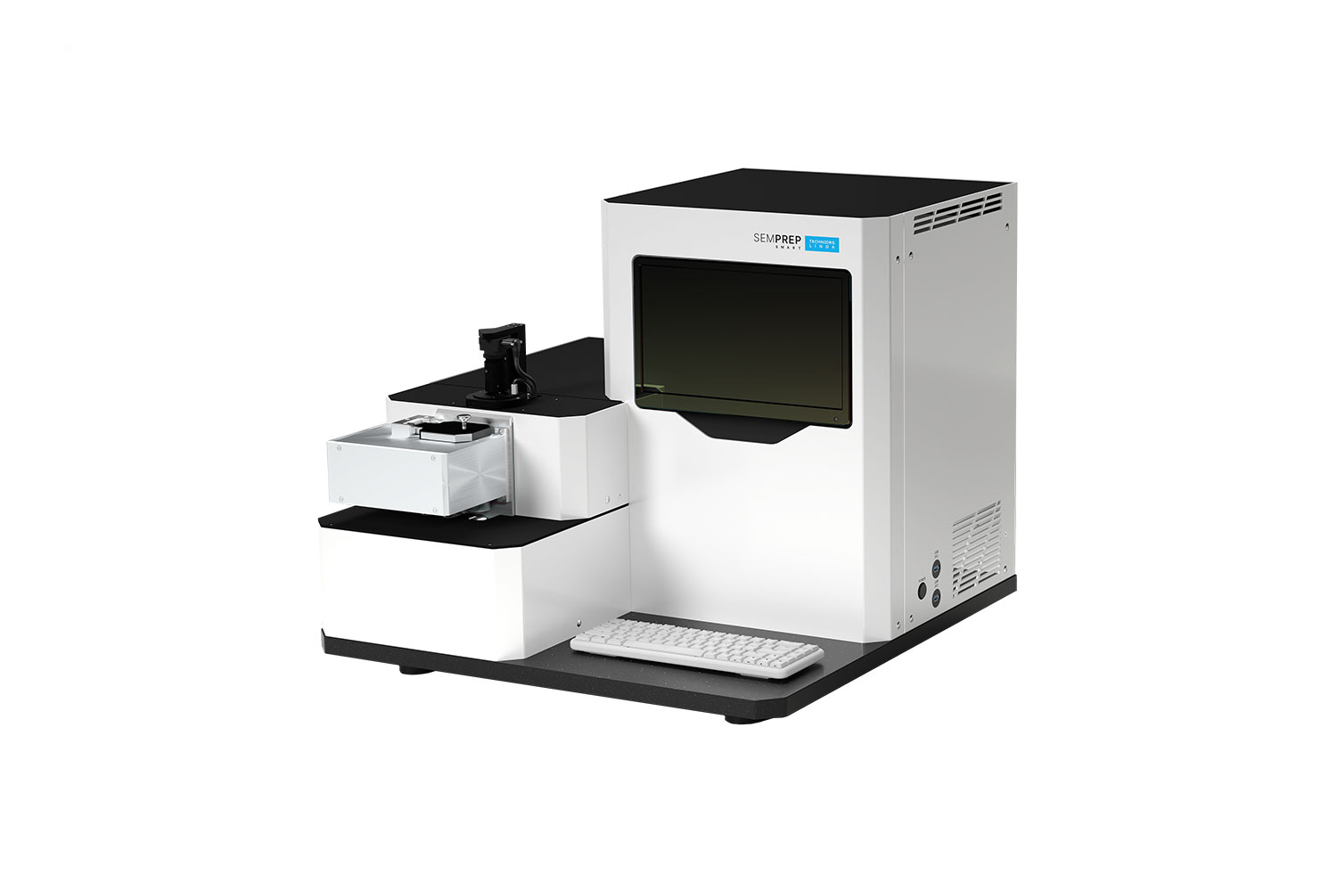
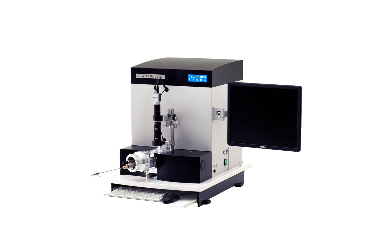
Ion milling is an essential part of the sample preparation process for electron microscopy. This method can remove surface contamination, planarize mechanically cross-sectioned samples, or thin and polish electron-transparent lamellae. These solutions are used across various fields like material science, biological research, and more.
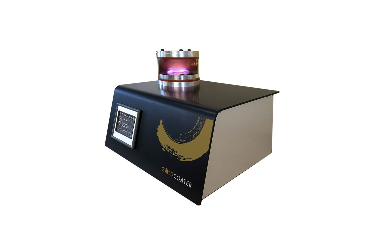
LUXOR sputter coaters are advanced devices that provide automated and controlled deposition of homogenous gold or platinum layers for use in reducing the effects of electric charging in scanning electron microscopy, offering improved image quality and reproducibility.
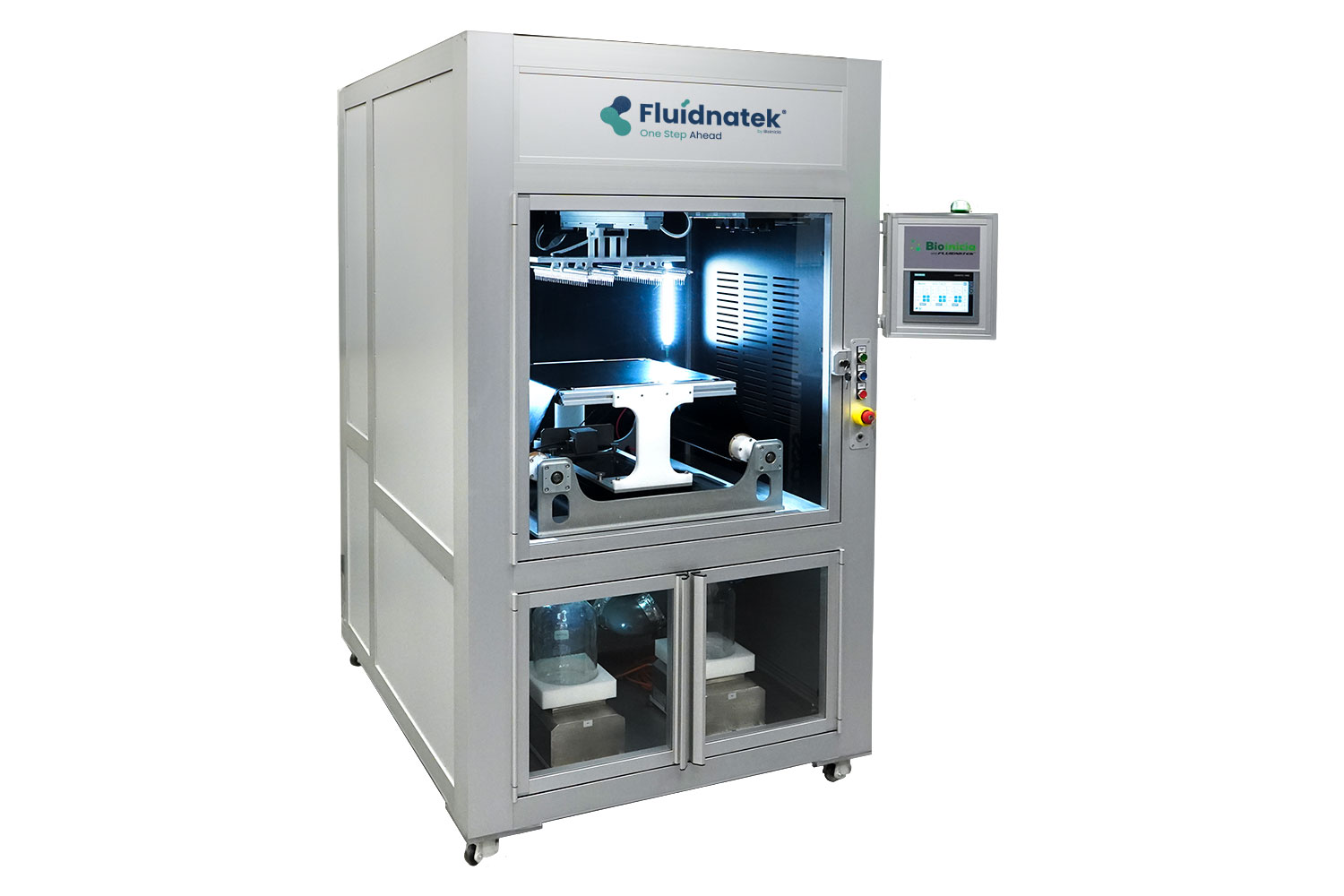
The Bioinicia family of instruments are designed to maximize control over electrospinning and electrospraying processes, enabling the reproducible fabrication of perfect nanofibers and nanoparticles.
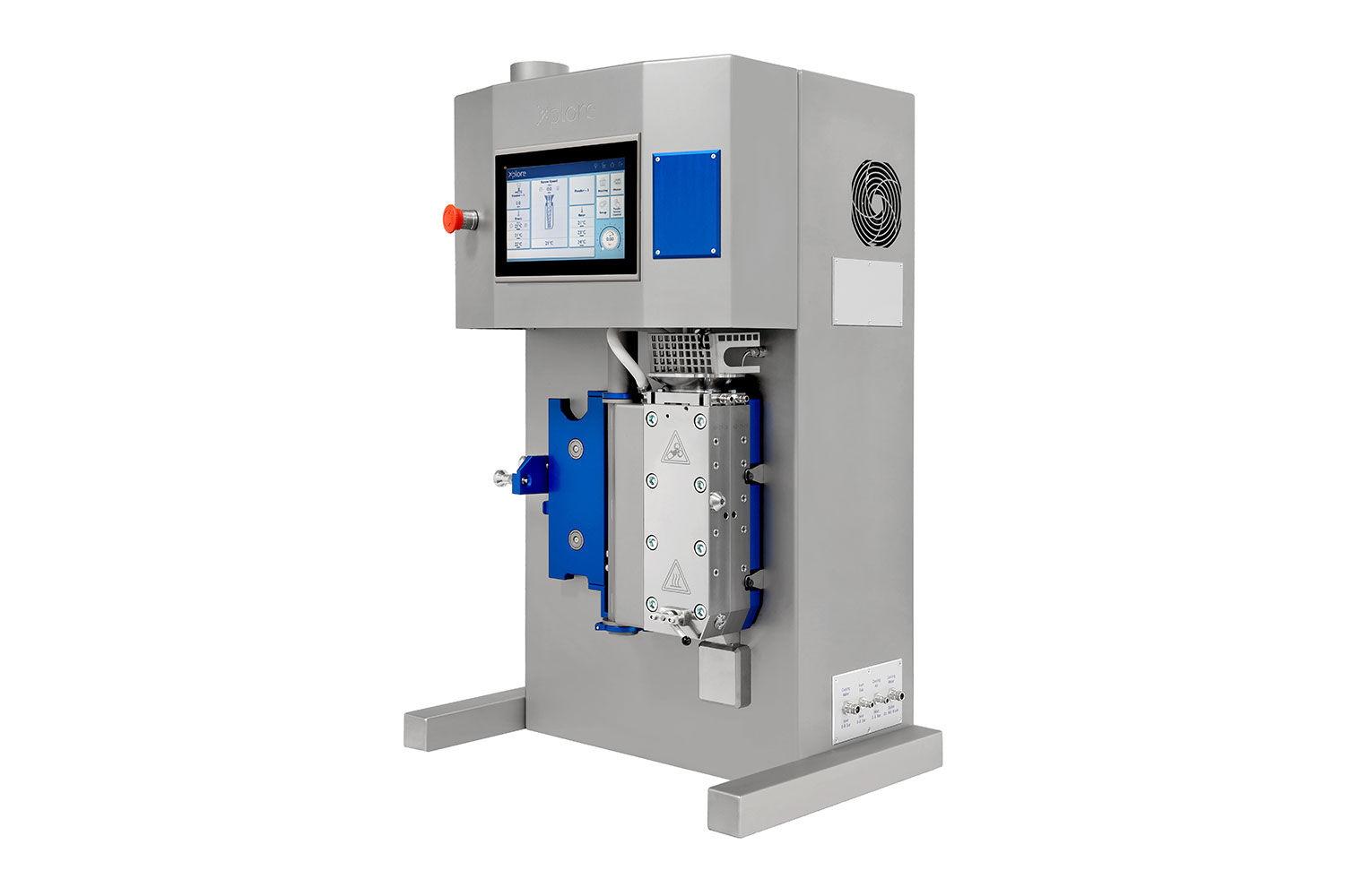
Xplore’s micro compounding and shaping instruments offer cost-effective solutions for R&D, accelerating time-to-market by replicating the performance of large twin-screw compounders.
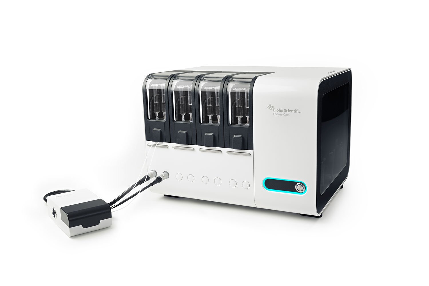
QSense QCM-D systems enable analysis of molecular interactions and surface properties in real-time. The QSense line allows vast exploration and experimentation, making it easy to find answers to research questions in various disciplines.
Attension tensiometers spans both optical and force characterization techniques, offering maximum versatility for studying surfaces and interfaces. The Theta line focuses on optical interfacial measurements, and the Sigma line is geared towards measurements of force.
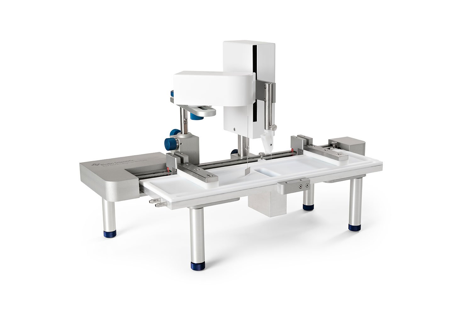
KSV NIMA has the widest selection of Langmuir and Langmuir-Blodgett Troughs of various sizes and functions. The modular trough systems are designed a frame capable of fitting multiple trough tops. The result is a flexible, and interchangeable solutions for multiple applications.
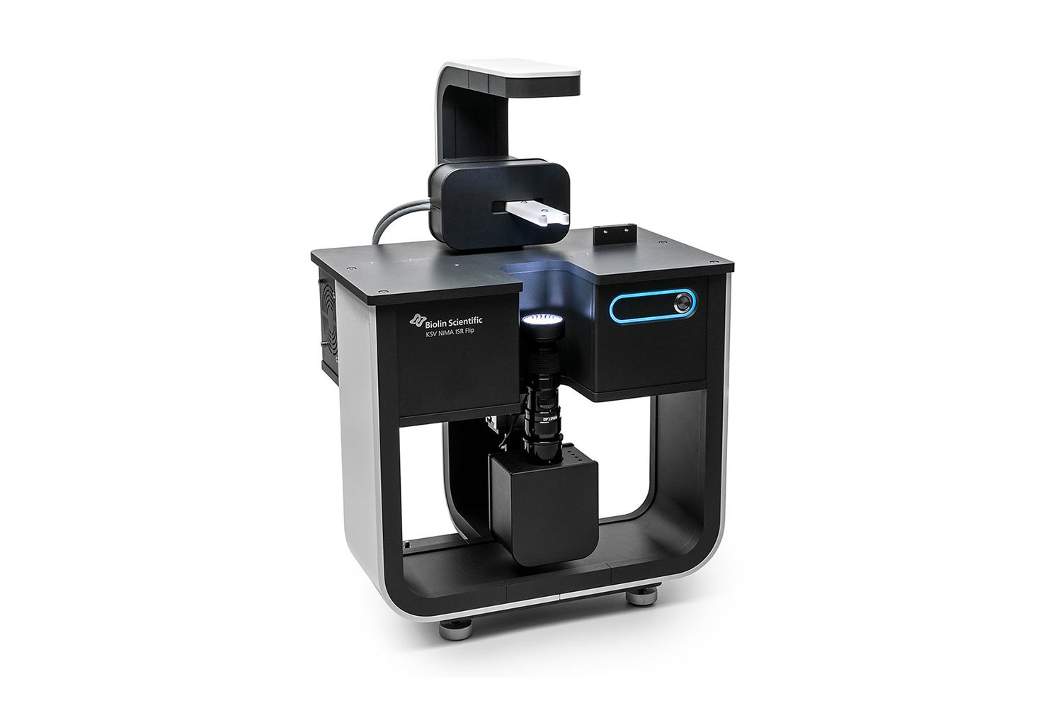
KSV NIMA ISR enables both dynamic and static measurements to define the viscoelasticity of the interfacial layers. With dynamic measurement, viscoelastic properties are measured as a function of frequency, time, strain, temperature or surface pressure.
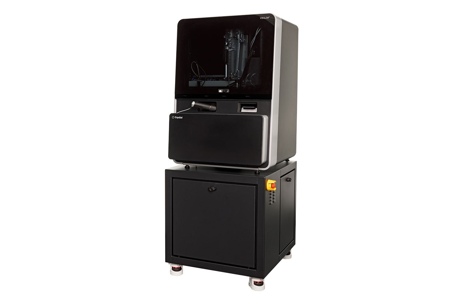
The VitroJet™ is designed to take the frustration and waste out of Cryo-EM sample preparation by allowing researchers to optimize the settings of ice quality, ice thickness and sample deposition before placement in the microscope.
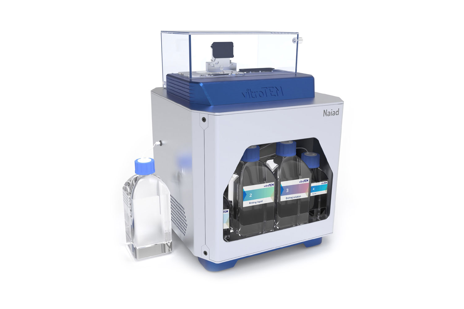
Graphene liquid cells (GLCs) are sub-micron sized pockets of liquid trapped between two layers of graphene. Naiad-1 makes working with GLCs easy and attainable for any laboratory, delivering efficient and reliable sample preparation.
Ion milling is an essential part of the sample preparation process for electron microscopy. This method can remove surface contamination, planarize mechanically cross-sectioned samples, or thin and polish electron-transparent lamellae. These solutions are used across various fields like material science, biological research, and more.