Tissue engineering is a progressively advancing field focusing on biological substitutes to improve or restore functions to damaged tissues or organs in the body. Many of these substitutes are found helping patients with burn wounds, diabetic wounds, diseased tissues, and many more. With such advancements, having a strong understanding of the tissue’s structures will help determine what the longevity is with these tissue substitutes.
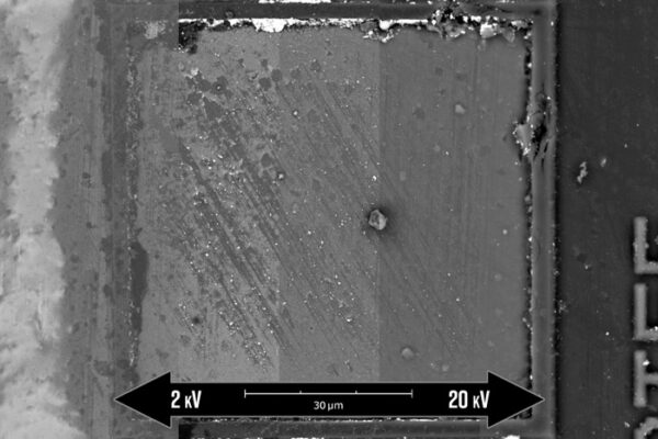
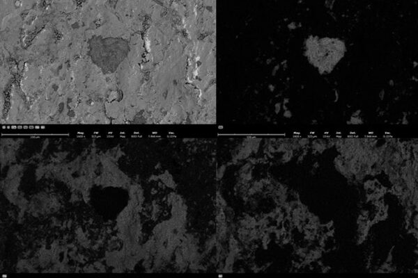
Scanning electron microscopy (SEM) is a valuable tool used in tissue engineering to help understand the structure and morphology of these tissues. Comparing the newly developed tissue with original or damaged tissue will aid scientists with identifying different parameters that they could adjust on solving their issues. The Phenom SEM is used to help identify many of these issues and make comparisons. Having access to different stages, such as the tensile stage, will help improve the outcome on producing data and contribute to the advancement of tissue engineering.