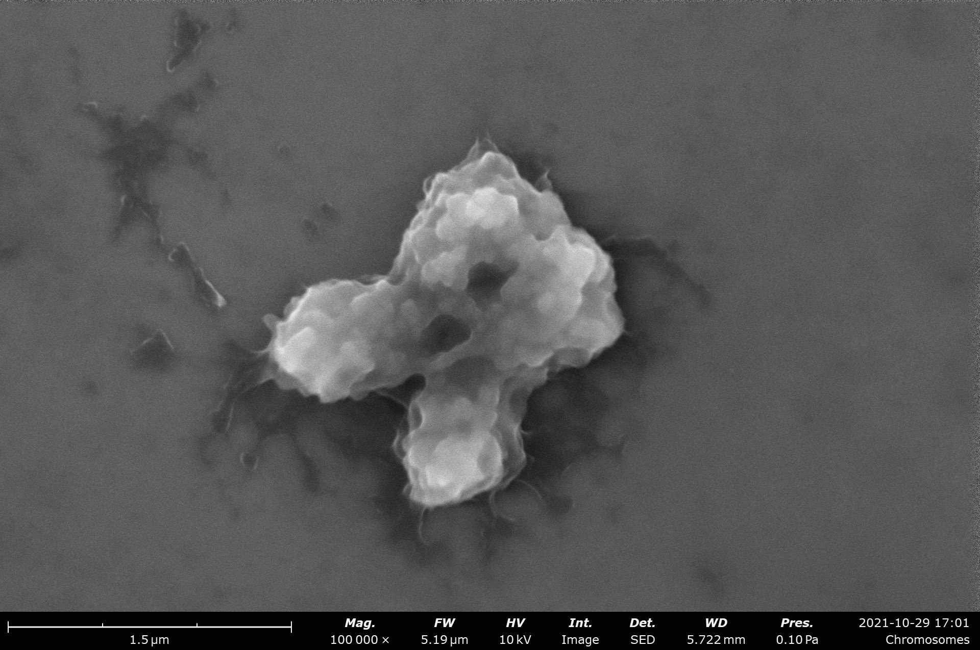
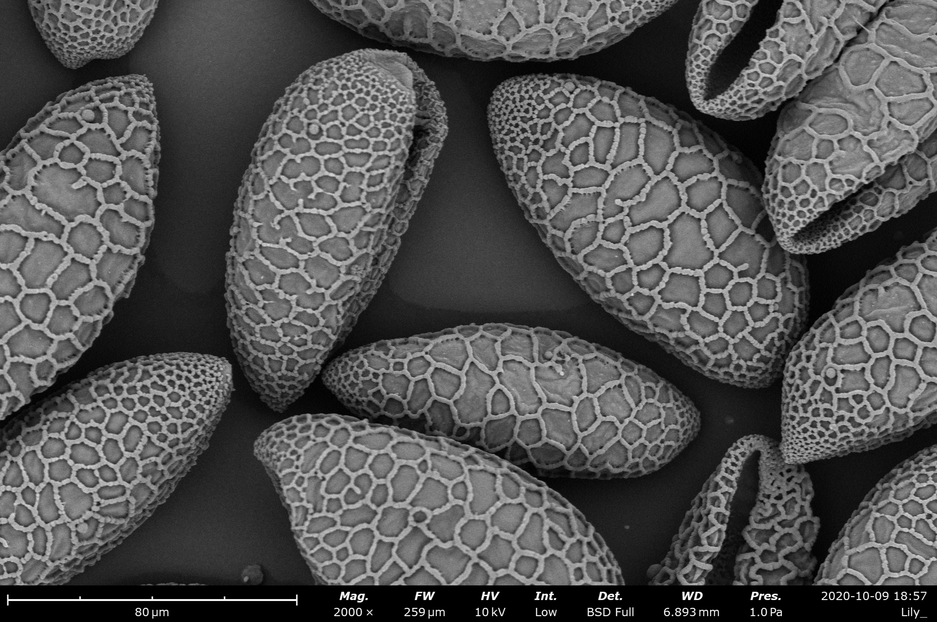
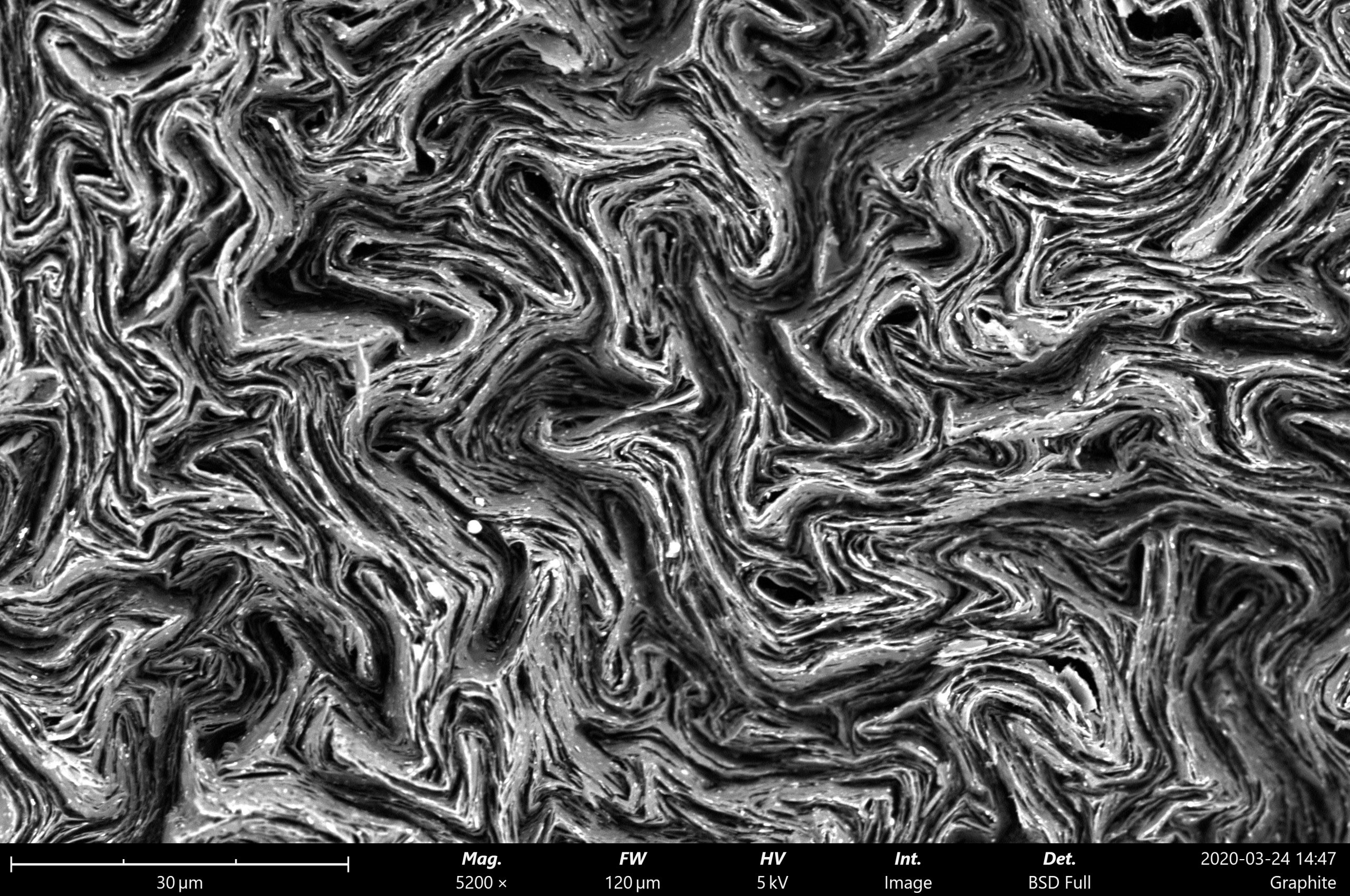
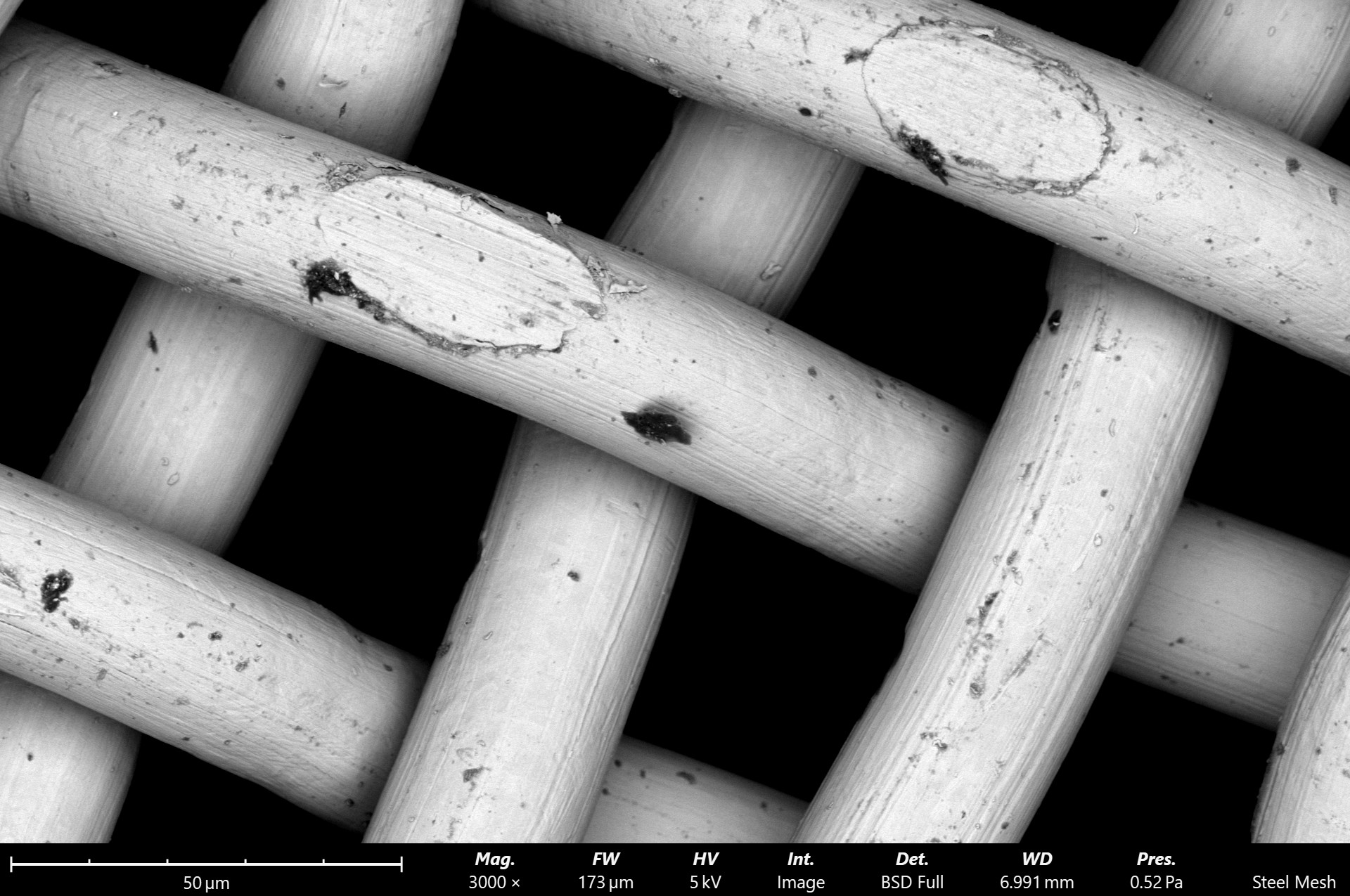
Scanning Electron Microscopy
Phenom Desktop SEMs
One of the great revolutions in electron microscopy was the miniaturization of scanning electron microscopes from large floor models to compact desktop models. Desktop SEMs, also called tabletop or benchtop SEMs, retain much of the power and capabilities of floor systems while also introducing a versatility of their own, fitting more easily into lab spaces and providing a suite of specialized software analysis tools. These systems prioritize ease of use and are ideal for in-house analysis at multi-user labs where everyone need not be an SEM specialist.
Speed to Data
High sensitivity detectors paired with the fastest vent/load cycle provides an average time-to-image of under 40 seconds
Intuitive
Operation
Imaging parameters and microscope functions controlled through a clear and navigable user interface
Low Cost of
Ownership
Resilient hardware enables low downtime and infrequent source changes, minimizing the overall maintenance cost
Download the Phenom SEM Product Brochure:
See available Configurations
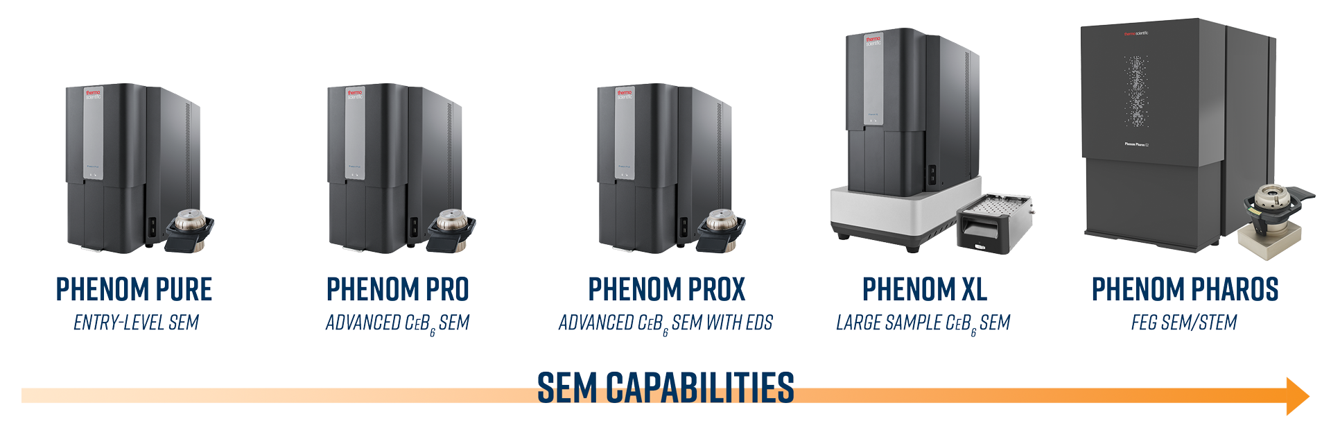
Phenom Desktop SEM Systems
The Phenom desktop scanning electron microscope from ThermoFisher was designed to be the easiest and most intuitive SEM ever built. Advanced electron sources in the form of cerium hexaboride crystals or field emission guns, along with multiple detectors, make it easy to acquire quality data and beautiful images. Imaging and analysis are mere clicks away!
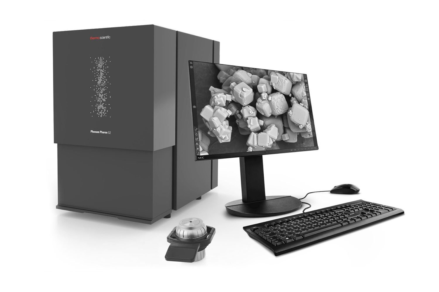
The Phenom Pharos is uniquely equipped with a field emission gun (FEG) and excels in applications demanding high spatial resolution. An unmatched coherent and bright electron beam, low kV accelerating voltage imaging, and automatic drift corrections make the Pharos a competitive alternative to many floor model SEMs.
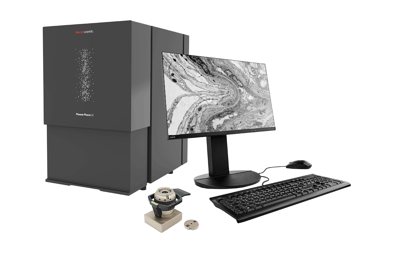
The Pharos STEM combines a high-brightness FEG source with a fully-integrated segmented STEM detector to generate STEM images with stunning contrast, even at low voltages. This compact desktop STEM-in-SEM system delivers the precision and resolution of nanoscale imaging traditionally reserved for larger, floor model systems.
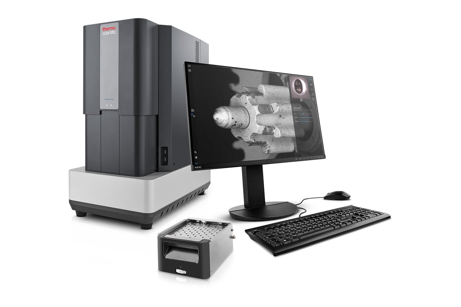
The Phenom XL has the largest sample capacity of all Phenom instruments, able to contain a high volume of small specimens, a single bulk mass, or anything in between. The XL also serves as the workhorse for an upgradeable automation software package known as ParticleX to push the boundaries of automated electron microscopy.
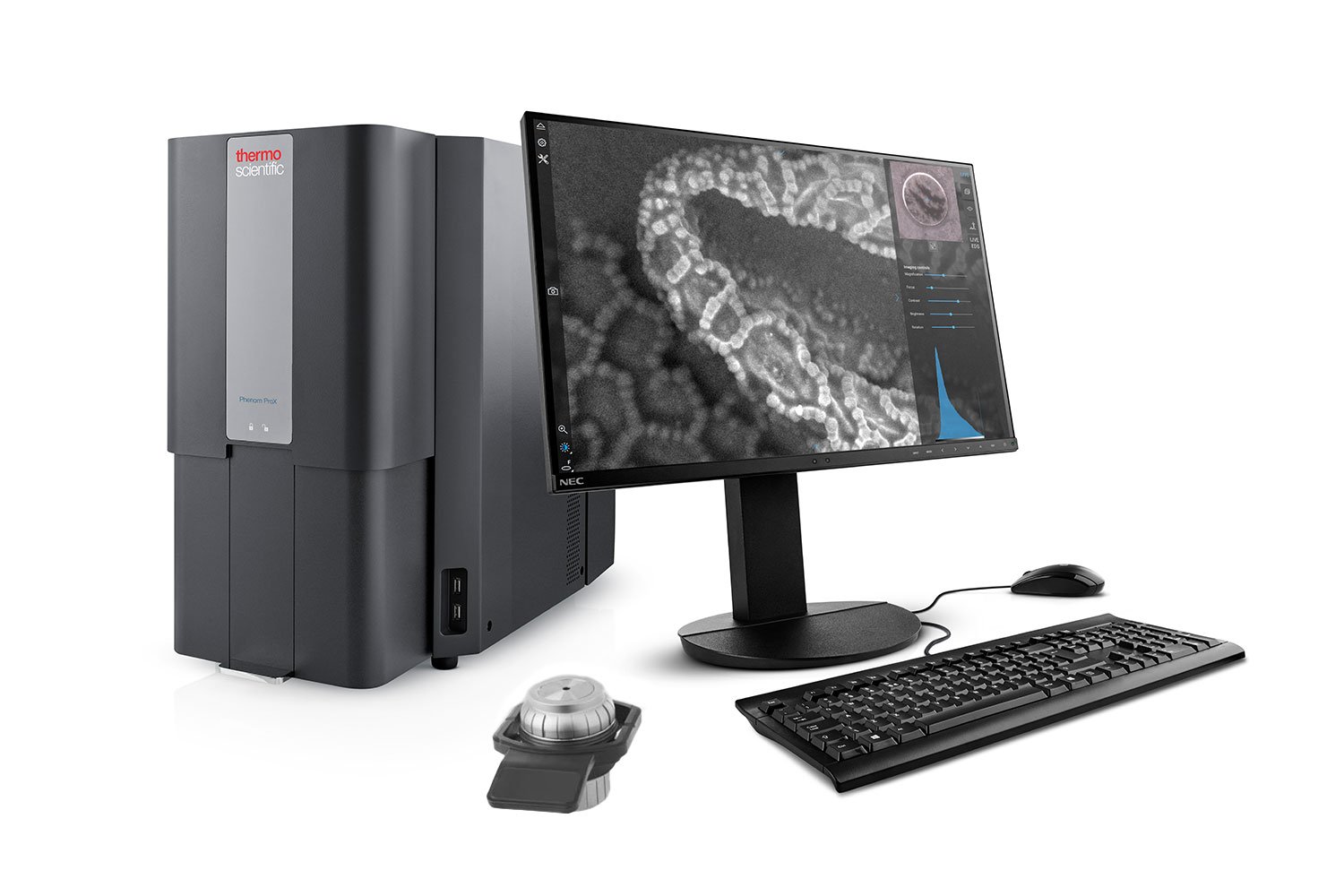
The Phenom Pro and ProX are the ultimate all-in-one SEM imaging systems, differentiated only by the presence of an integrated energy dispersive X-ray spectroscopy (EDS) detector. The unique and powerful core architecture characteristic of the Phenom line combines with a host of data analysis features to provide the most complete desktop SEM solution on the market.
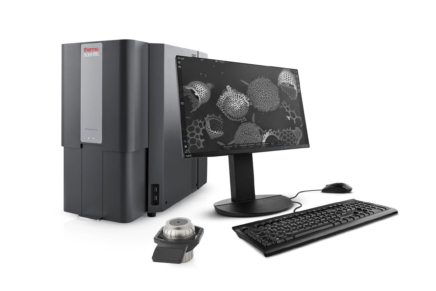
The Phenom Pure is an entry-level, economical desktop SEM useful for many different applications, whether it be in teaching environments, simple R&D, or for general imaging. The Pure provides a straightforward imaging solution to visualize sample features, surfaces, and structures and is a user-friendly tool to seamlessly transition from light optical to electron microscopy.
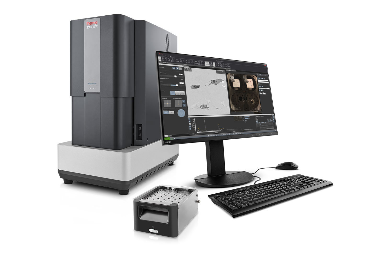
An empowered model of the Phenom XL, ParticleX is a powerful quality control tool with automated SEM-EDS workflows for targeted, unbiased data analysis. Packages include: Technical Cleanliness, Steel, Battery, Gunshot Residue, and Additive Manufacturing; all of which comply with industry-relevant reporting standards like ASTM E45 (Steel) and ASTM E1588 (GSR)
Model: |
Phenom Pharos |
Phenom XL |
Phenom Pro/ProX | Phenom Pure |
|---|---|---|---|---|
Specifications |
||||
| Electron Source | Field Emission Gun (FEG) | CeB6 crystal | CeB6 crystal | CeB6 crystal |
| Max. SEM Magnification | 2,000,000x | 200,000x | 350,000x | 175,000x |
| SEM Resolution – BSD | 3 nm | 9 nm | 8 nm | 15 nm |
| SEM Resolution – SED | <2 nm | 9 nm | 6 nm | 15 nm |
| Acceleration Voltages | 1 – 20 kV | 1.5 – 20 kV | 1.5 – 20 kV | 2, 5 or 10 kV |
| Navigation Camera | ||||
| Sample Handling | Single pin stub 25 mm diameter |
Up to 36 pin stubs 100 mm x 100 mm |
Single pin stub 25 mm diameter |
Single pin stub 25 mm diameter |
| Detectors | BSD – Four-quadrant backscattered electron detector (standard) SED – Everhart-Thornley secondary electron detector (optional) EDS/EDX – Integrated energy dispersive x-ray spectrometer (optional) |
|||
| STEM Detector | ||||
Knowledgebase
SEM Knowledgebase
Webinar
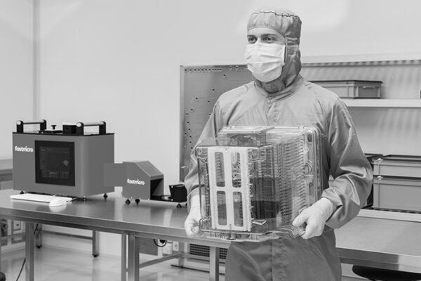
Accelerating Surface Particle Contamination Control by Combining Fastmicro and Phenom Technologies
Surface particle contamination remains a critical challenge across advanced manufacturing…
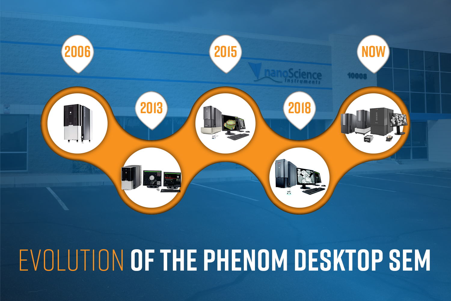
Evolution of the Phenom Desktop SEM
The Phenom Scanning Electron Microscope (SEM) is turning 20 this year! In celebration of t…
White Paper
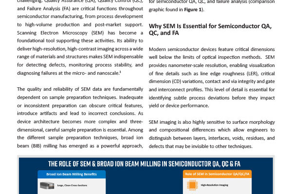
SEM & BIB milling for QA, QC, and Failure Analysis in Semiconductor Devices
Introduction As semiconductor devices continue to scale in complexity and shrink in featur…