Phenom Scanning Electron Microscope
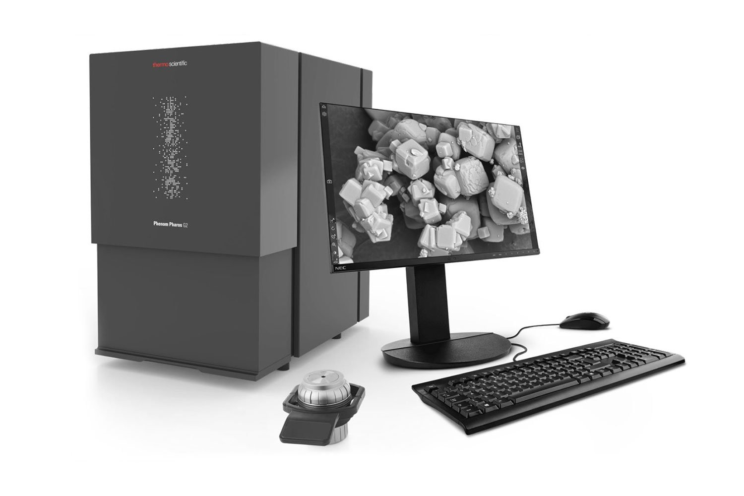
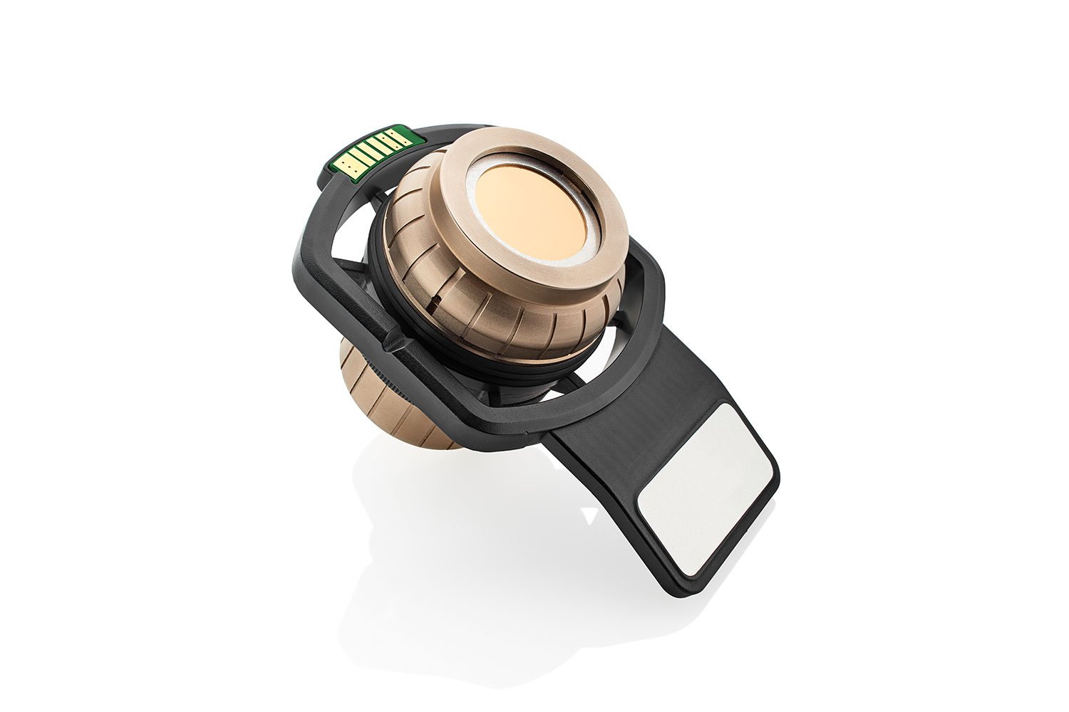
Phenom Pharos Desktop SEM
The Phenom Pharos Desktop SEM uses a field emission gun (FEG) as its source of electrons for applications that demand the highest resolution. Benefitting from a broad accelerating voltage range of 1-20 kV, the Pharos can accommodate a variety of insulating and beam-sensitive samples with low kV imaging, an ability enhanced further by a temperature-controlled sample holder, and scanning transmission mode.
High-Resolution Imaging
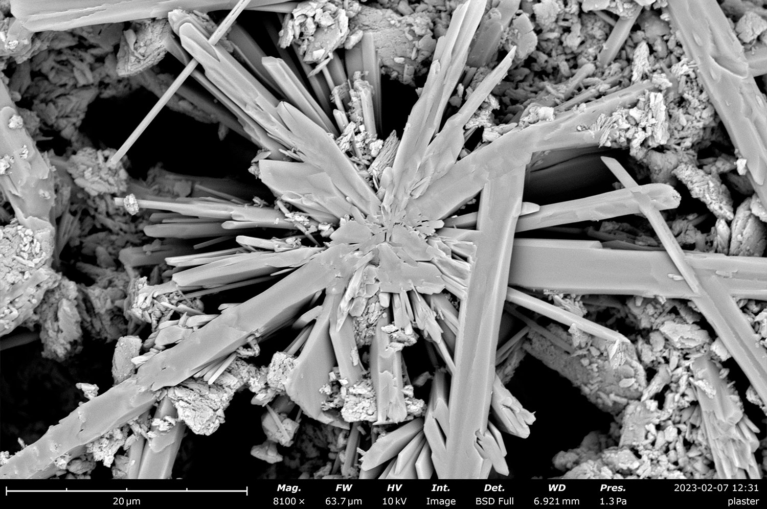
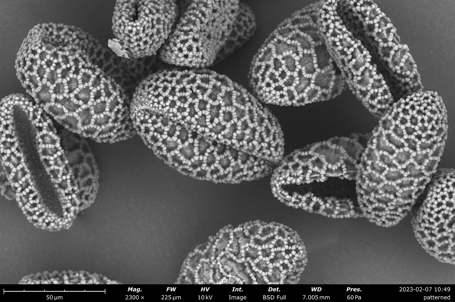
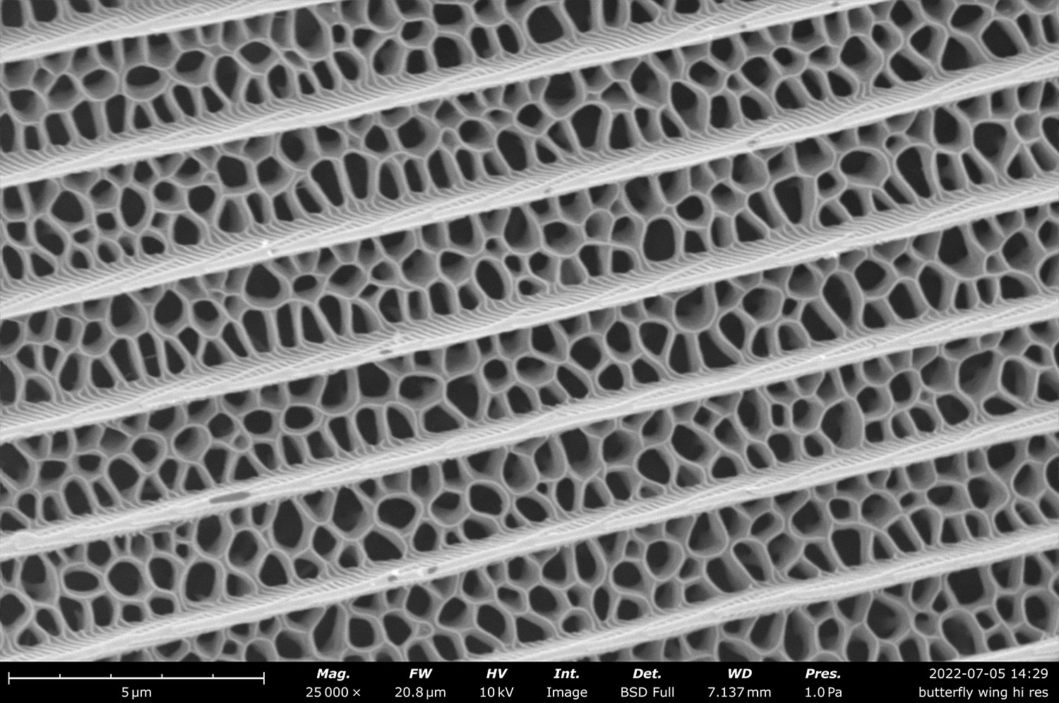
Experience unparalleled imaging performance with field electron emission, delivering the highest resolution among desktop SEMs.
Low Voltage Mode
Achieve precise imaging of insulating and beam-sensitive materials with accelerating voltages as low as 1 kV.
Upgradeable to STEM
Effortlessly switch to transmission imaging with the integrated plug-and-play STEM detector and sample holder.
Talk to an Instrumentation Specialist Today!
The Only Tabletop SEM with a Field Emission Source
The Phenom Pharos G2 Desktop SEM is the premier field emission desktop scanning electron microscope (SEM), from ThermoFisher, designed to be the most accessible FEG system with the smallest footprint on the market, yet also capable of delivering the highest resolution images of any other desktop SEM.
The core architecture of the Pharos is of the same design that has established the Phenom line of desktop SEMs as the world’s best- selling desktop SEMs: a segmented backscattered electron detector (BSD), optional secondary electron detector (SED), energy-dispersive X-ray detector (EDS), and scanning transmission electron microscopy (STEM) detector, low vacuum modes, and sophisticated suite of analytical software permit a thorough analysis of any sample. Coupling the core characteristics of Phenom SEMs with the low kV imaging and field emission technology of the Pharos pushes the boundaries of electron microscopy to new limits by enabling the detailed characterization of sensitive samples in pristine states.
Model: |
Phenom Pharos |
Phenom XL |
Phenom Pro/ProX | Phenom Pure |
|---|---|---|---|---|
Specifications |
||||
| Electron Source | Field Emission Gun (FEG) | CeB6 crystal | CeB6 crystal | CeB6 crystal |
| Max. SEM Magnification | 2,000,000x | 200,000x | 350,000x | 175,000x |
| SEM Resolution – BSD | 3 nm | 9 nm | 8 nm | 15 nm |
| SEM Resolution – SED | <2 nm | 9 nm | 6 nm | 15 nm |
| Acceleration Voltages | 1 – 20 kV | 1.5 – 20 kV | 1.5 – 20 kV | 2, 5 or 10 kV |
| Navigation Camera | ||||
| Sample Handling | Single pin stub 25 mm diameter |
Up to 36 pin stubs 100 mm x 100 mm |
Single pin stub 25 mm diameter |
Single pin stub 25 mm diameter |
| Detectors | BSD – Four-quadrant backscattered electron detector (standard) SED – Everhart-Thornley secondary electron detector (optional) EDS/EDX – Integrated energy dispersive x-ray spectrometer (optional) |
|||
| STEM Detector | ||||
Phenom Pharos Desktop SEM
Product Features
Field emission gun
The high brightness of a Schottky field emission gun (FEG) brings superb resolution across an accelerating voltage range of 1- 20 kV.
Charge Reduction Mode
Image non-conducting samples without extra sample preparation or gold coating using the built-in low vacuum mode. No special detectors or additional infrastructure are required.
Low Voltage Imaging
The ability to perform SEM imaging at low accelerating voltages has several advantages, from increasing surface sensitivity to reducing charging and sample damage.
Proven Ease-of-Use Operation
Live SEM images, an optical overview, clean menus, and straightforward controls culminate into an accessible and intuitive, friendly user experience.
Flexible Integration & Minimal Footprint
Phenom Desktop SEMs are designed to deliver the capabilities of larger SEMs in a more compact and versatile form. Their streamlined design allows them to fit seamlessly into any workspace, whether on a surface or tabletop. With built-in vibrational stability, the Phenom offers a highly durable and robust SEM solution suitable for a wide range of applications.
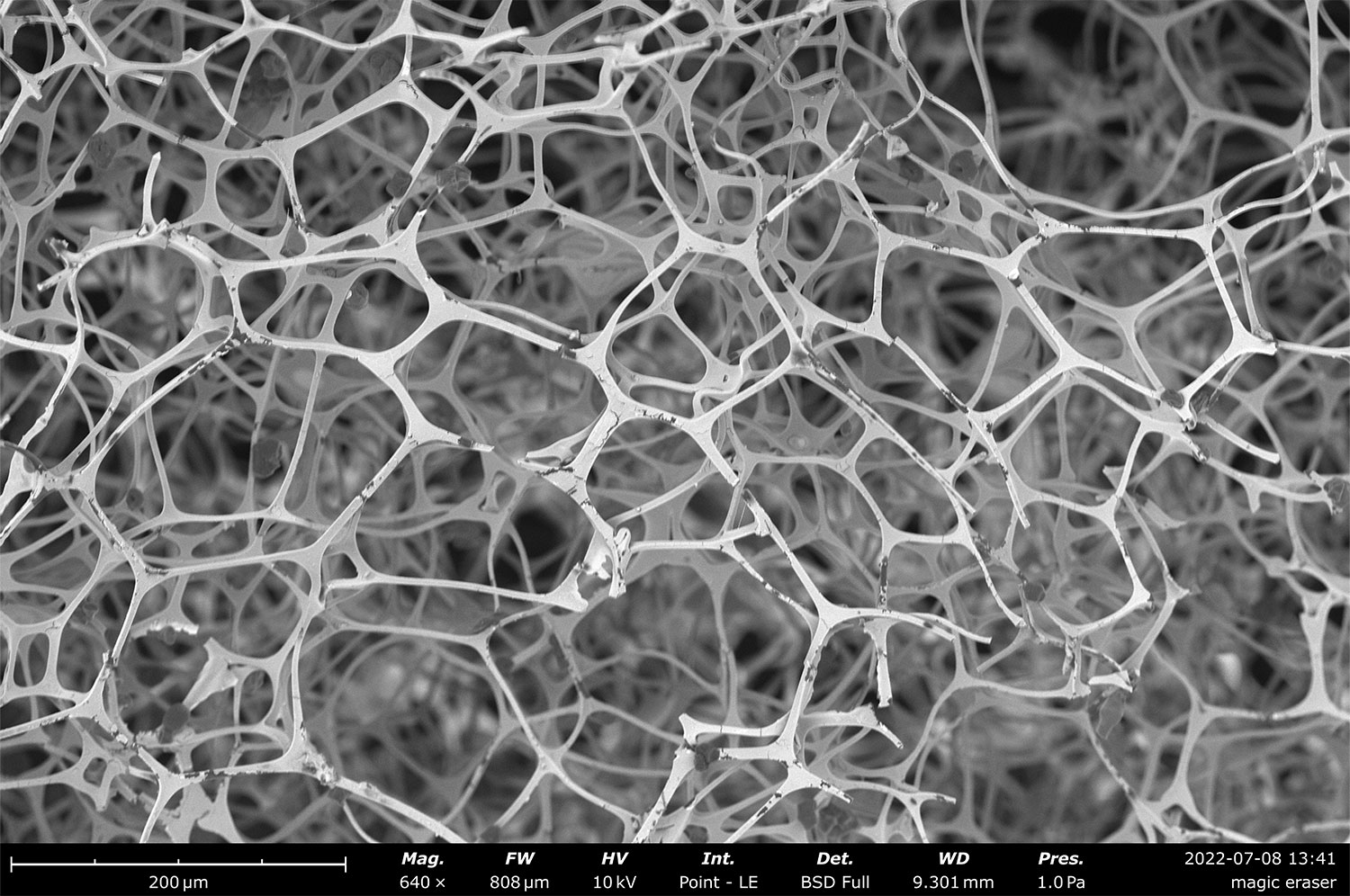
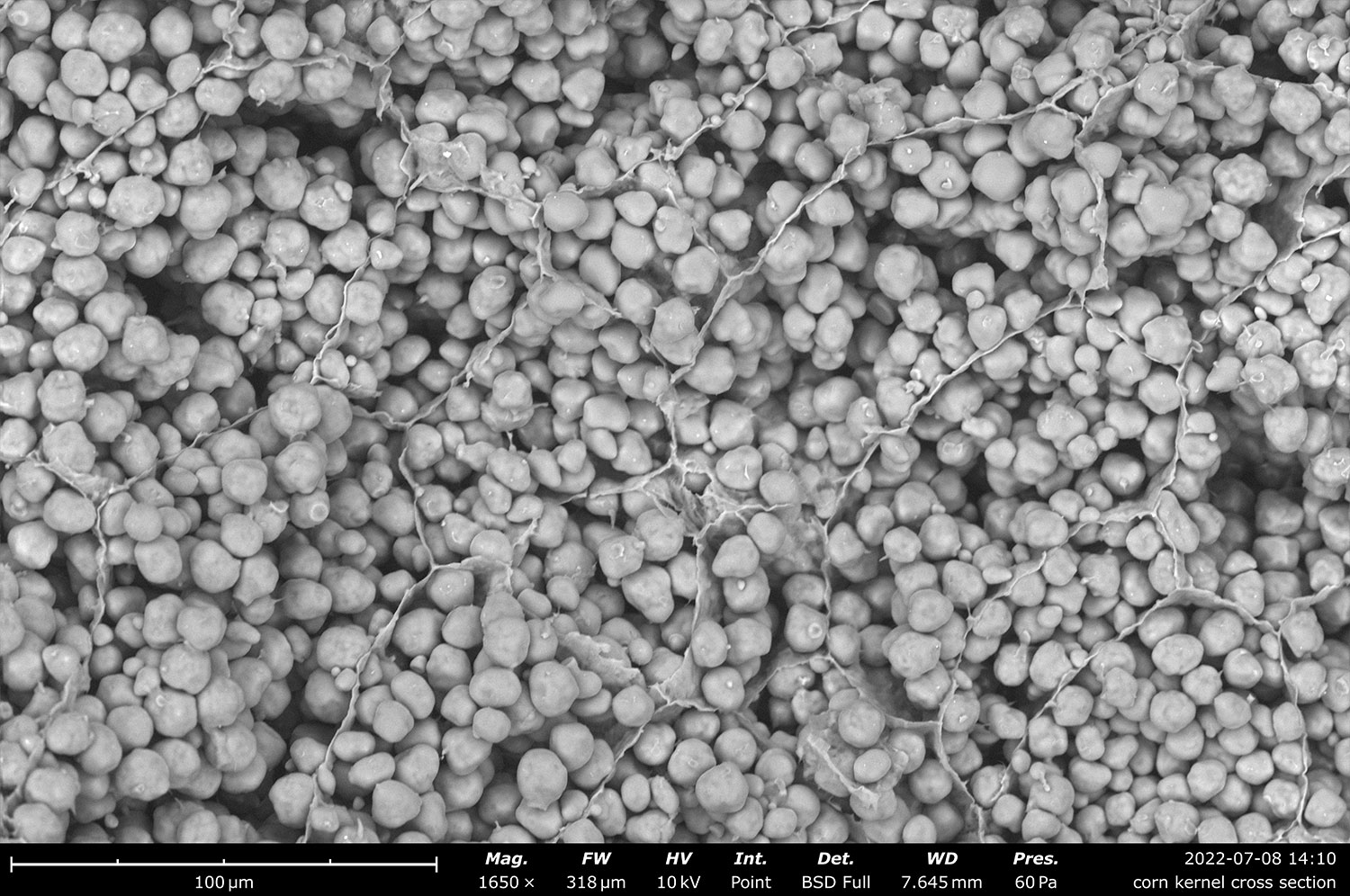
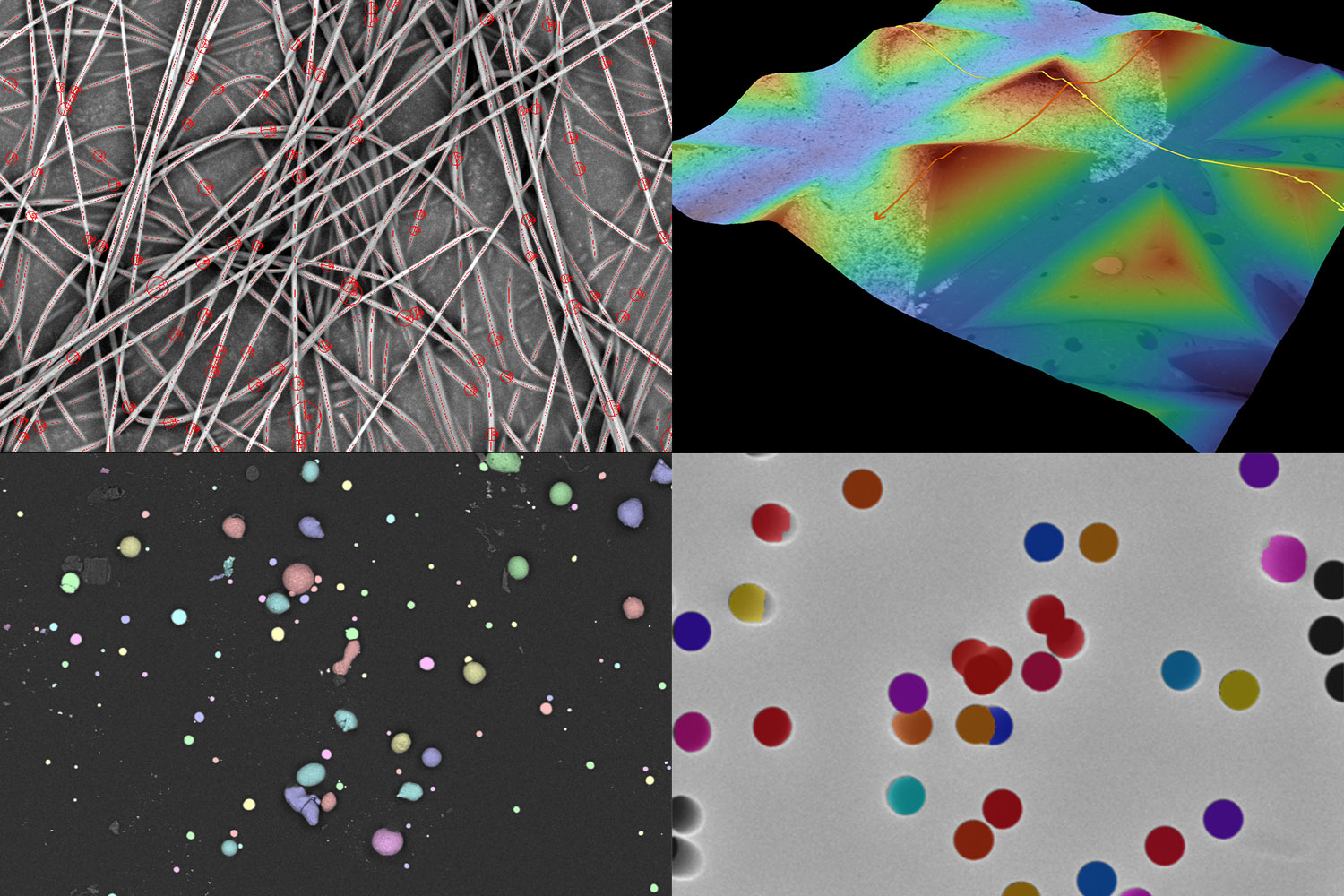
Four-Segment Backscattered Electron Detector (BSD)
The four-segmented backscattered electron detector (BSD) delivers sharp compositional contrast between light and heavy elements. Included as a standard feature in every Phenom system, it also offers topographic modes for qualitative visualization of surface roughness, enhancing surface detail and material differentiation.
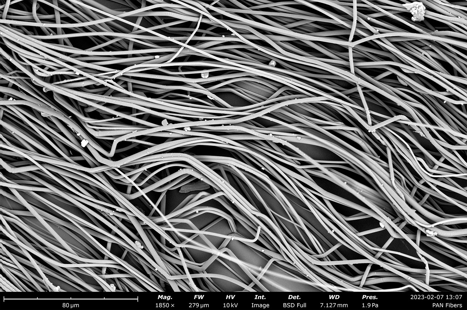
Secondary Electron Detector (SED)
Secondary Electron Detectors (SED) are perfect for high-resolution, surface-sensitive SEM imaging, providing detailed topographical information through secondary electron emissions. This allows for crisp visualization of fine surface features. With an SED, Phenom users can also activate a mixed mode, combining both SED and BSD signals at a customizable ratio for comprehensive imaging analysis.
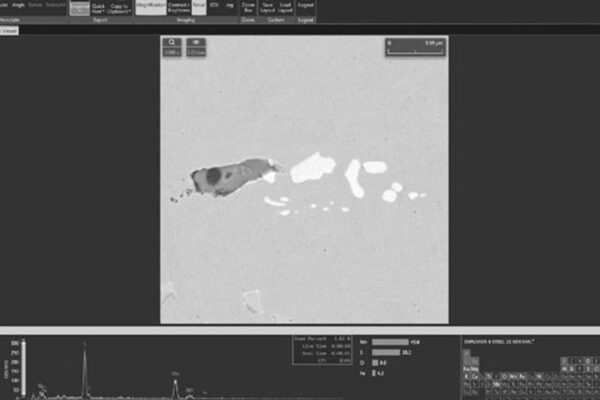
Energy Dispersive X-ray Spectroscopy Detector (EDS)
Energy Dispersive X-Ray Spectroscopy (EDS) detectors are fully integrated into Phenom SEMs, enabling efficient quantitative surface chemistry analysis. EDS provides rapid and precise results across multiple analysis modes, including regions, points, and lines, making it a versatile tool for a wide range of applications.
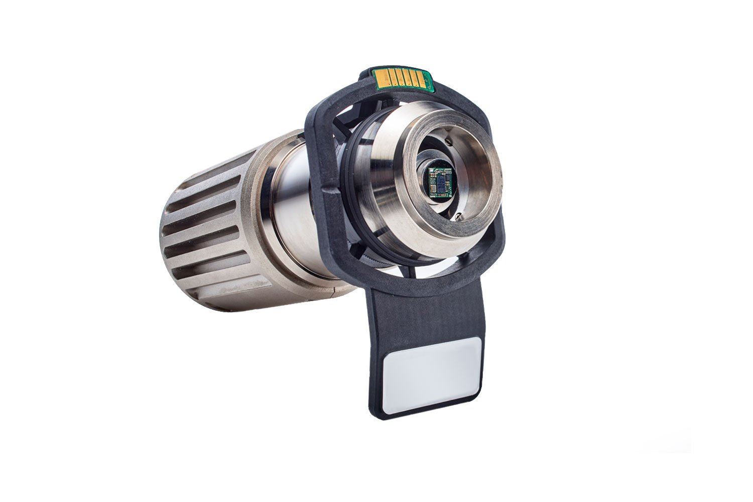
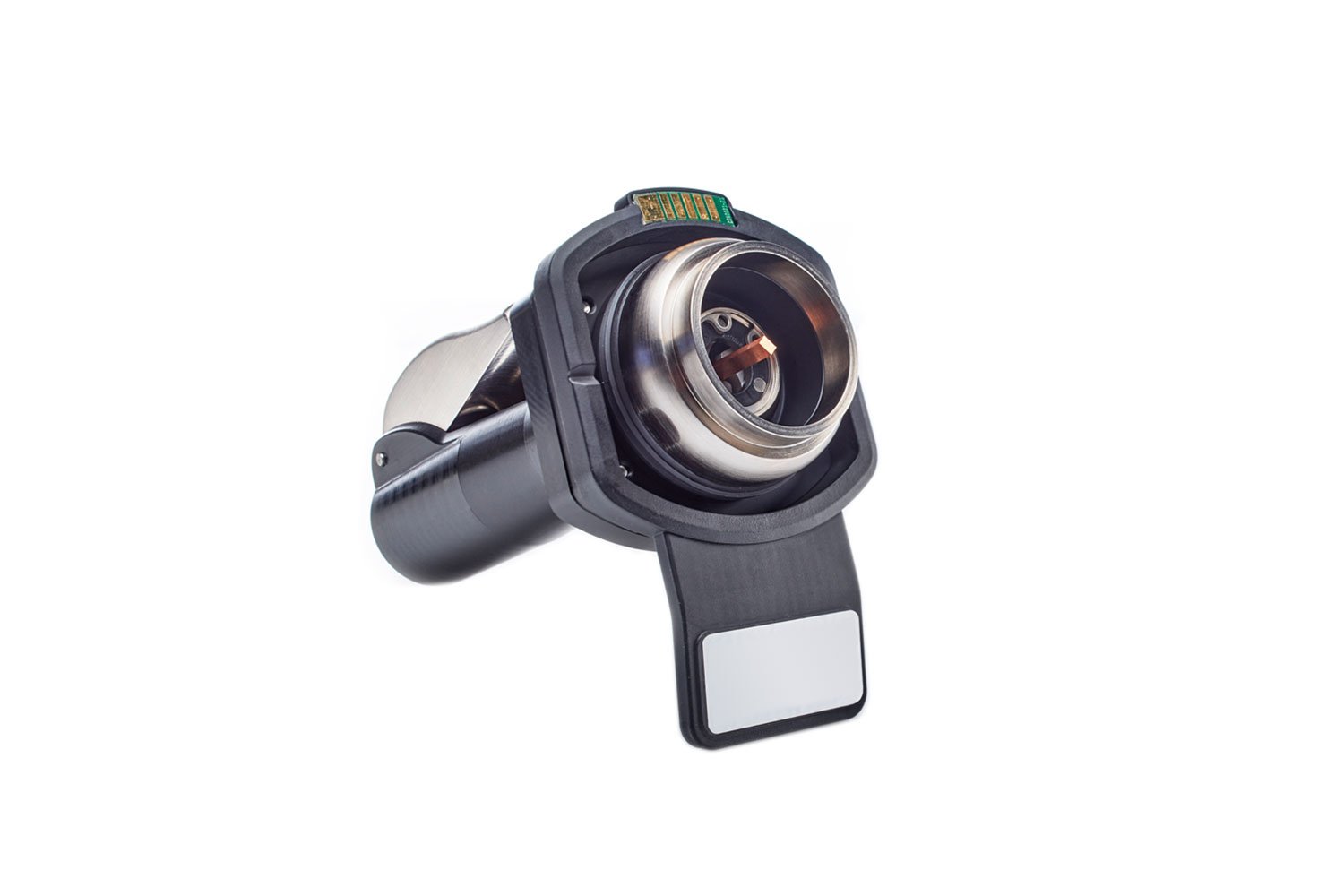
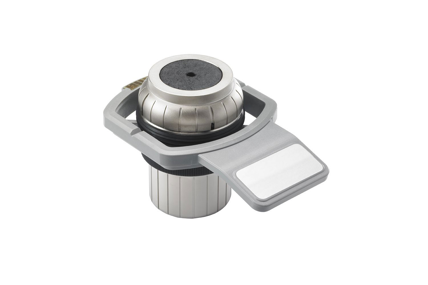
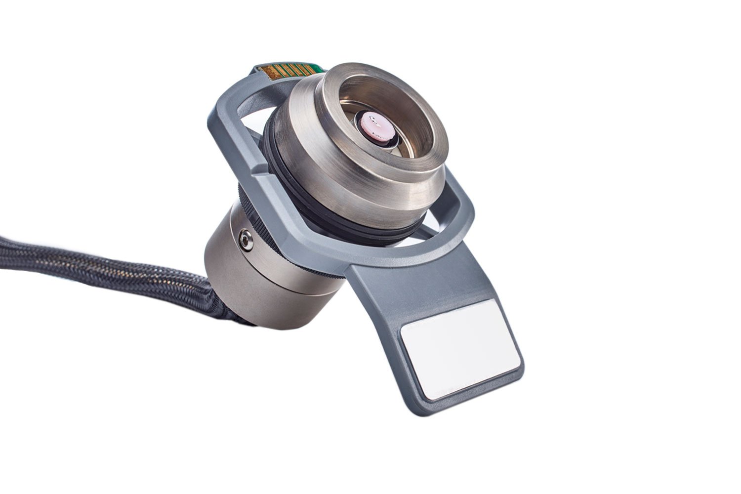
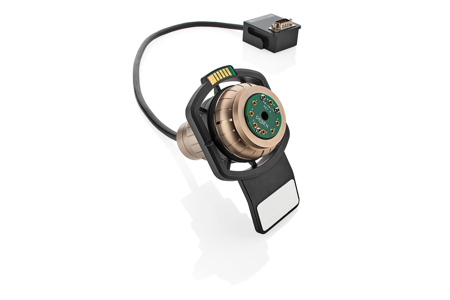
Pharos STEM Detector & Sample Holder
With the Phenom Pharos STEM detector, transmission imaging becomes more accessible, efficient, and cost-effective. This holder seamlessly adds functionality with its plug-and-play design and intuitive workflow. Switch between SEM and STEM modes, adjust imaging parameters, and acquire high-quality images with just a few clicks.
Phenom Pharos Desktop SEM
Accessories
Motorized Tilt & Rotate Sample Holder
Revealing the hidden features of samples is made possible with the Motorized Tilt & Rotation sample holder. This holder allows for angular tilting and rotation of the sample stage, allowing for imaging of the same area from multiple angles and improved visualization of three-dimensional structures.
Microtool Sample Holder
The Microtool Sample Holder features an iris style clamp, specially designed for securely holding axially shaped objects or samples with a high aspect ratio. Ideal for observing drill bits, milling tools, and injection needles, simply secure the sample with a non-destructive clamping mechanism and start imaging within minutes.
Charge Reduction Sample Holder
The Charge Reduction Sample Holders effectively reduces electric charge buildup, minimizing the amount of preparation needed to analyze nonconductive samples. Paper, polymers, organic materials, ceramics, glass, and coatings are but a few of the materials accommodated by these holders.
Temperature Controlled Sample Holder
The Temperature Controlled Sample Holder protects sensitive specimens and enables imaging of hydrated samples by Peltier cooling and heating. With a temperature range of -25°C to 50°C, samples can be maintained at sub-zero temperatures to prevent damage from the vacuum and electron beam.
Electrical Feedthrough Sample Holder
The Electrical Feedthrough Sample Holder enables in-situ electrical measurements. The holder has six quick release pins that can be used to measure or apply voltages and currents to the sample while it is under the SEM column. This holder is ideal for observation of MEMS devices and for performing failure analysis.
Metallurgical Sample Holder
The Metallurgical Sample Holder is designed to support resin-embedded samples. It comes in two variations – standard and charge reduction, with the option for two sizes of inserts. This holder speeds up SEM analysis for metallurgical samples.
Phenom Pharos Desktop SEM
Software
Phenom Prosuite Software
ProSuite Software, developed specifically for Phenom SEMs, enhances data throughput and eliminates user bias from manual measurements, ensuring more accurate and reliable results. It can extract actionable data on the size and shape of features in SEM images.
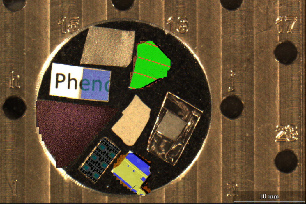
MAPS
MAPS software is a transformative tool for Phenom Desktop Scanning Electron Microscopes (SEMs), designed to boost both the efficiency and depth of your analytical workflows. MAPS features seamless image stitching – delivering high-resolution SEM images and EDS maps over expansive sample areas. MAPS also facilitates multi-modal correlative analysis across different analytical systems.
ChemiSEM / ChemiPhase
Streamline elemental and phase analysis with integrated data collection and advanced analysis capabilities. ChemiSEM provides real-time elemental mapping during live imaging and ChemiPhase transforms EDS maps into color-coded phase maps.
Phenom Pharos Desktop SEM
SEM Sample Preparation
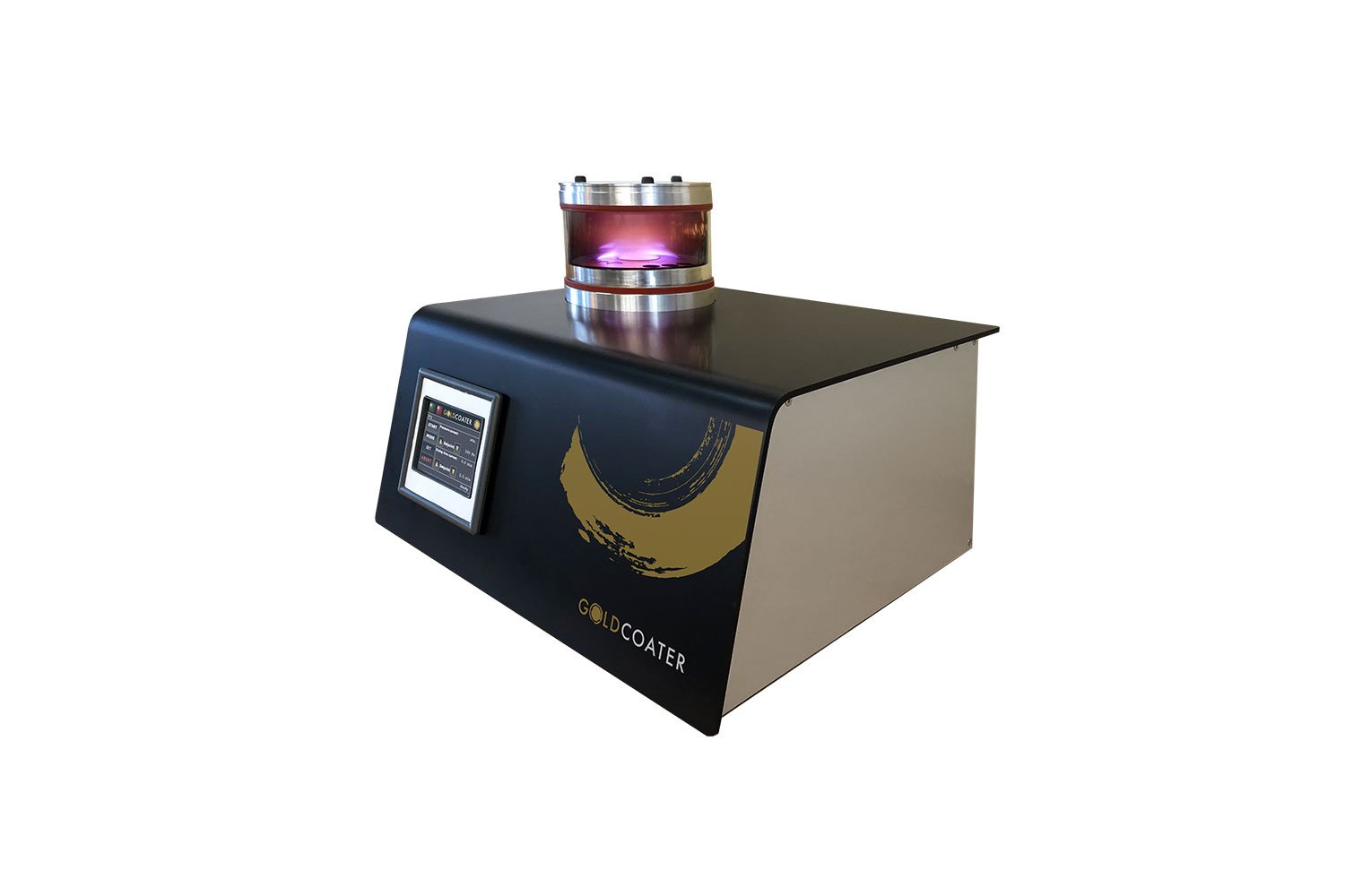
Sputter Coaters
Minimize electric charging and enhance SEM image quality with uniform gold and platinum coatings.
Ion Mills
Prepare artifact-free samples for high quality cross-sectional analysis. Especially useful for EBSD and failure analysis.
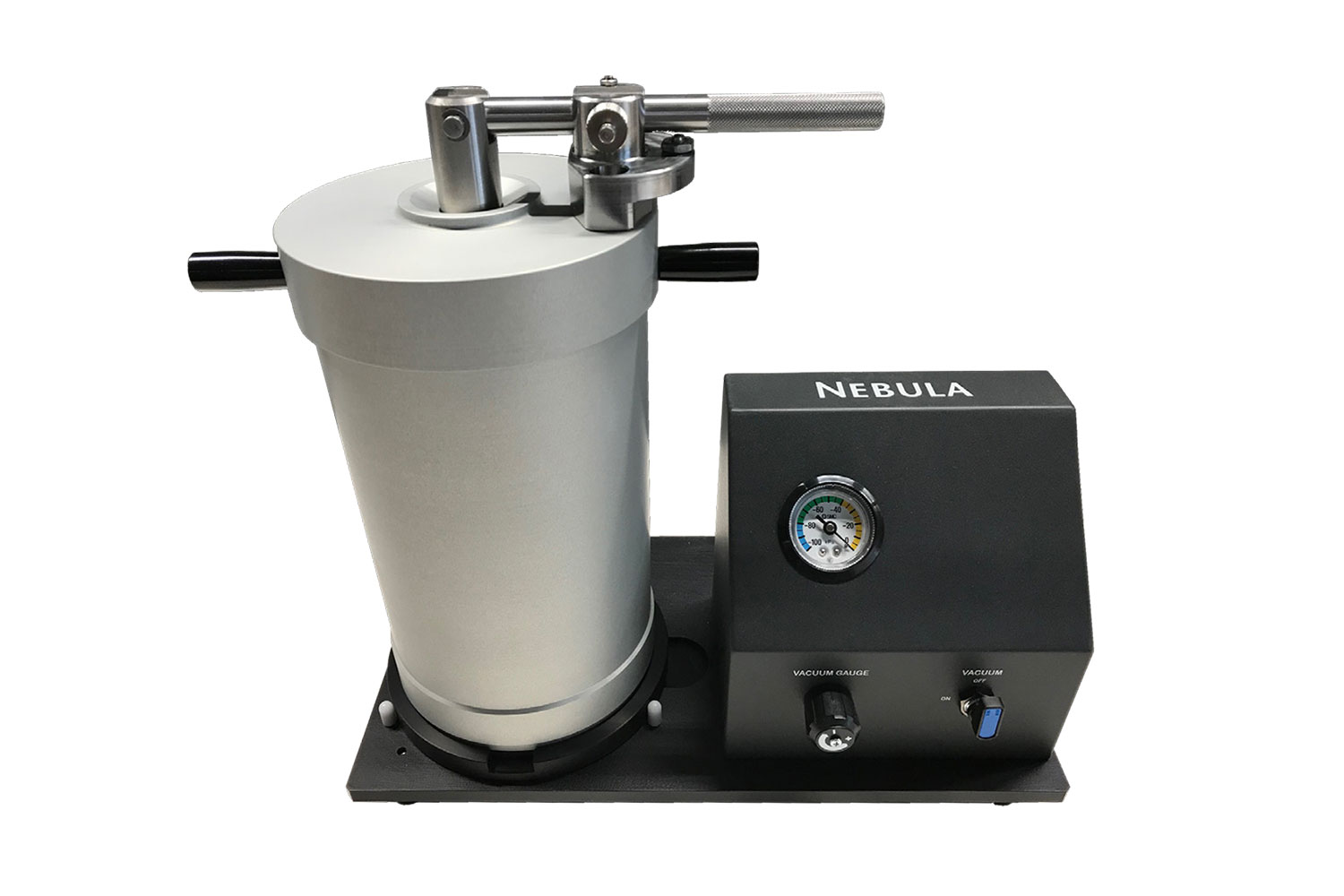
Nebula Particle Disperser
The Nebula is designed to assist in dispersing powder samples, facilitating their preparation for SEM analysis.
Phenom Pharos Desktop SEM
Product Knowledgebase
Faster, Smarter Inclusion Analysis for Modern Metallurgical Labs
From bearings that are used in the satellites to the specialty alloy metals used in orthop…
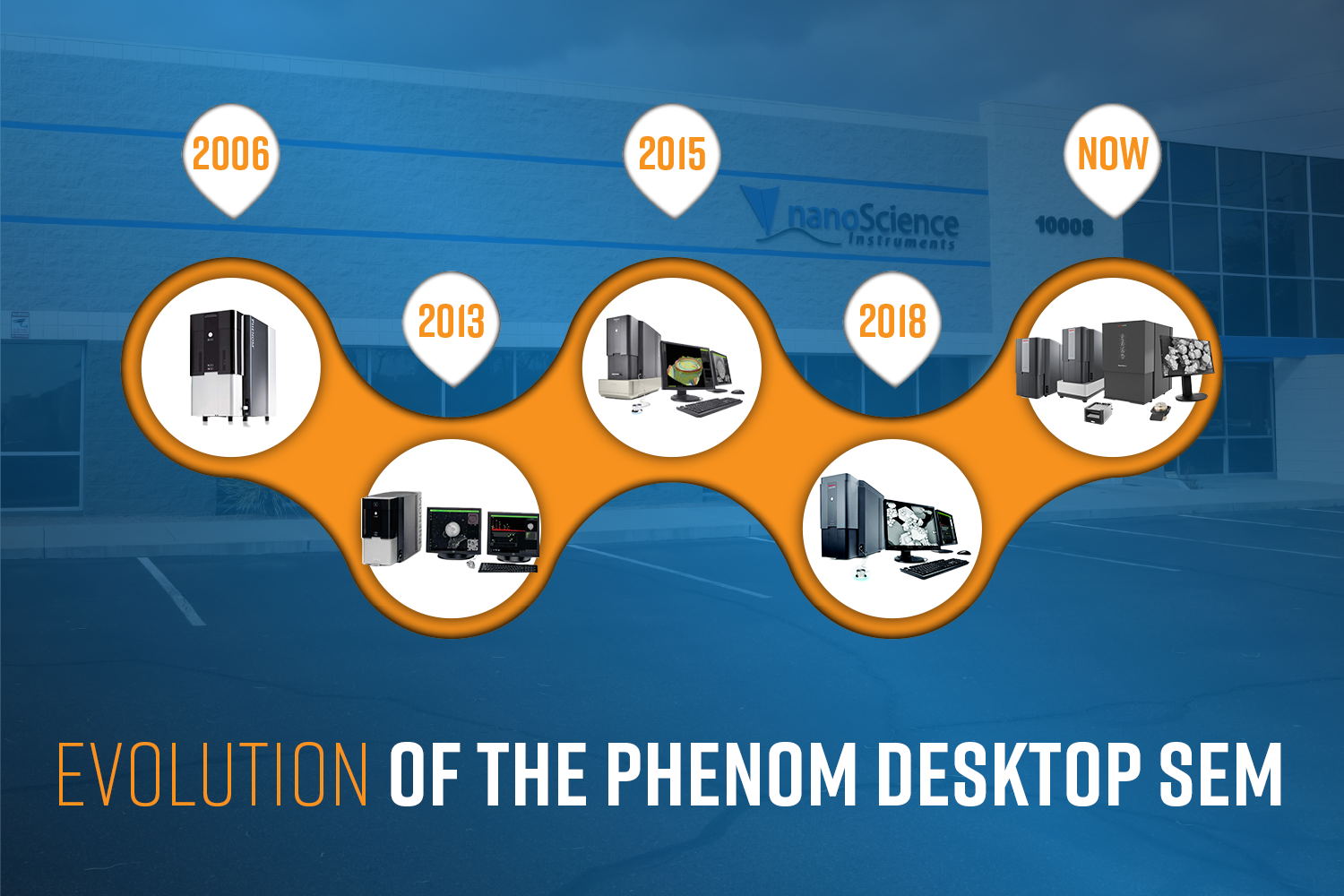
Evolution of the Phenom Desktop SEM
The Phenom Scanning Electron Microscope (SEM) is turning 20 this year! In celebration of t…
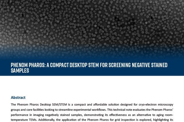
Phenom Pharos: A Compact Desktop STEM for Screening Negative Stained Samples
The Phenom Pharos Desktop SEM/STEM is a compact and affordable solution designed for cryo-…